. 2022 Feb 11;17(2):e0262328.
doi: 10.1371/journal.pone.0262328. eCollection 2022.

Prickle isoform participation in distinct polarization events in the Drosophila eye

Affiliations

Prickle isoform participation in distinct polarization events in the Drosophila eye

Bomsoo Cho et al. PLoS One. .

Abstract

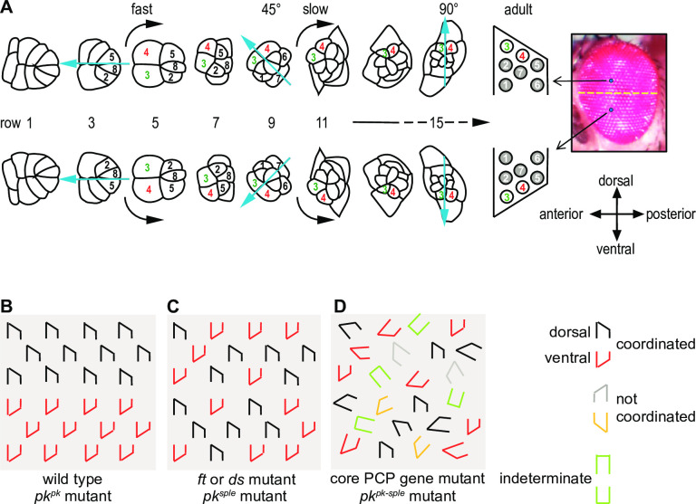
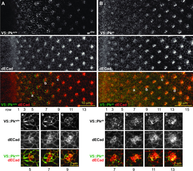
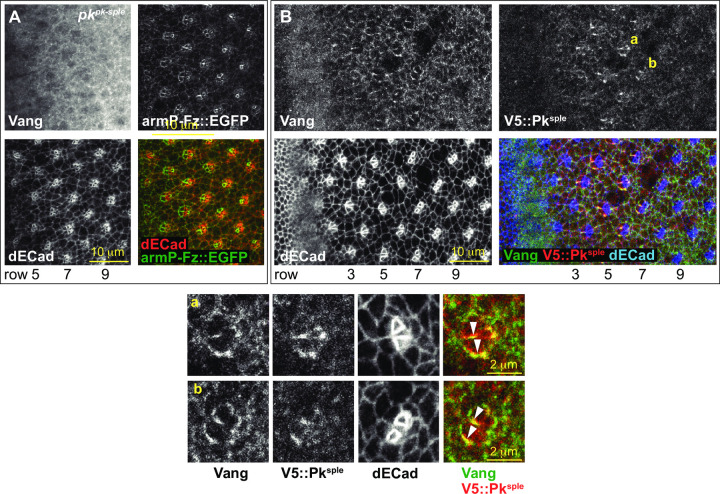
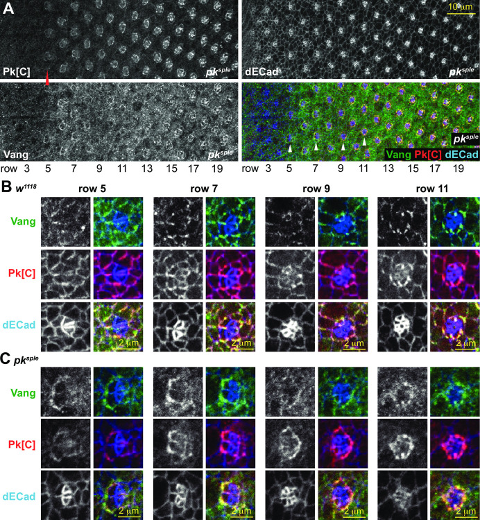
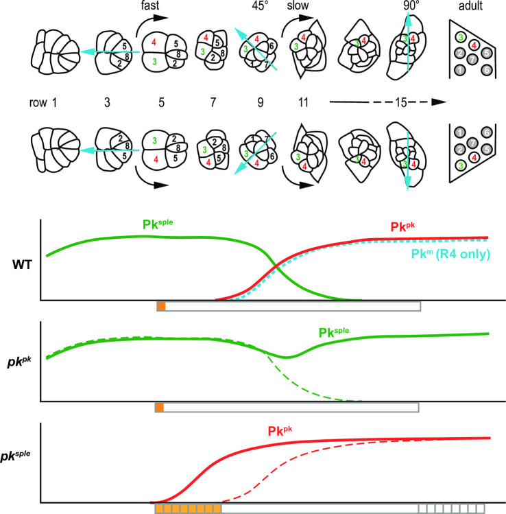
Planar cell polarity (PCP) signaling regulates several polarization events during development of ommatidia in the Drosophila eye, including directing chirality by polarizing a cell fate choice and determining the direction and extent of ommatidial rotation. The pksple isoform of the PCP protein Prickle is known to participate in the R3/R4 cell fate decision, but the control of other polarization events and the potential contributions of the three Pk isoforms have not been clarified. Here, by characterizing expression and subcellular localization of individual isoforms together with re-analyzing isoform specific phenotypes, we show that the R3/R4 fate decision, its coordination with rotation direction, and completion of rotation to a final ±90° rotation angle are separable polarization decisions with distinct Pk isoform requirements and contributions. Both pksple and pkpk can enforce robust R3/R4 fate decisions, but only pksple can correctly orient them along the dorsal-ventral axis. In contrast, pksple and pkpk can fully and interchangeably sustain coordination of rotation direction and rotation to completion. We propose that expression dynamics and competitive interactions determine isoform participation in these processes. We propose that the selective requirement for pksple to orient the R3/R4 decision and their interchangeability for coordination and completion of rotation reflects their previously described differential interaction with the Fat/Dachsous system which is known to be required for orientation of R3/R4 decisions but not for coordination or completion of rotation.


Conflict of interest statement

The authors have declared that no competing interests exist.

Figures

Fig 1
Fig 1. Ommatidial maturation and PCP mutant phenotypes.
A. Cells are recruited into photoreceptor and other cell fates stereotypically as ommatidia develop. Photoreceptor cell numbers are shown. Simultaneously, the clusters rotate in opposite directions on either side of the equator in an initial fast and then a slow phase. Each row is approximately two hours older than the subsequent row. B. Schematic of ommatidia in a wildtype and a pkpk mutant eye. Ommatidia have opposite chirality in the dorsal and ventral hemispheres. C. In ft, ds or pksple mutant eyes, dorsal and ventral type ommatidia are intermixed and no equator is apparent. D. In core PCP mutant eyes, including pkpk-sple eyes, some ommatidia fail to distinguish R3 and R4 (indeterminate), dorsal and ventral type ommatidia are intermixed, and ommatidia rotate in the correct or incorrect direction and to varying extent, or not at all.
Fig 2
Fig 2. Pkpk and Pksple apico-lateral junctional expression in wildtype eyes.
A. Endogenously tagged V5::Pksple stained for V5 (green in merge) and dECad (red in merge), showing V5::Pksple enriched at apico-lateral junctions of early ommatidia. Aa-c. Enlarged images of ommatidia from rows 5, 7 and 9 as marked. B. Endogenously tagged V5::Pkpk stained for V5 (green in merge) and dECad (red in merge), showing V5::Pkpk enriched at apico-lateral junctions of late ommatidia. Ba-d. Enlarged images of the ommatidia from rows 7, 9, 11 and 13 as marked.
Fig 3
Fig 3. The relationship between Vang apico-lateral localization and Pk.
A. In a pkpk-sple mutant eye, Vang fails to localize at apico-lateral junctions. B. Vang co-localizes with V5::Pksple at apico-lateral junctions of early ommatidia. Vang (green in merge), V5::Pksple (stained for V5; red in merge) and dECad (blue in merge). Ba-b. Enlarged images of ommatidia from panel B rows 6 and 7 as marked, showing co-localization and asymmetry of Vang and V5::Pksple at the equatorial sides of R3 and R4 (arrowheads).
Fig 4
Fig 4. Vang and Pk in a pksple mutant eye.
A. A pksple mutant eye stained with the Pk[C] common antibody (detects mostly Pkpk) and a Vang antibody. Note that Pkpk is weakly detected at apico-lateral junctions beginning at row 5, somewhat earlier than in control (w1118), and Vang is detected in a similar pattern. Enlarged images of ommatidia from a control (w1118; B) and a pksple (C) mutant eye from rows 5, 7, 9 and 11 as marked in A and S3 Fig (for B). Vang appears at apico-lateral junctions somewhat later, concomitant with expression of Pkpk, in the pksple mutant eye. Vang (green in merge), Pk[C] (red in merge) and dECad (blue in merge). Asymmetry of Pkpk and Vang localization is often delayed, as in this row 7 ommatidium.
Fig 5
Fig 5. Delayed restoration of vang or knockdown of pkpk.
Expression of control wRNAi, vang, or pkRNAi was driven by GMR-GAL4 in mutant or wild type backgrounds as indicated. GMR-GAL4 expression becomes significant by row 7 as judged by loss of Pk[C] signal upon GMR-GAL4-driven knockdown by pkRNAi (S6 Fig). Restoration of vang expression corrects most indeterminate R3/R4 decisions, and allows most rotations to complete to 90°, but rotation direction is uncoordinated with the R3/R4 decision. In a pksple mutant eye, intermixed dorsal and ventral ommatidia rotate to 90°, but delayed knockdown of remaining pk isoforms (mostly pkpk) causes under-rotation of some ommatidia.
Fig 6
Fig 6. Pksple replaces Pkpk in apico-lateral domains of older pkpk mutant ommatidia.
A. A pkpk mutant eye stained with anti-Pksple and the common Pk[C] antibody. The common antibody recognizes Pksple and Pkm in this condition. Pksple is detected in apico-lateral junctions of early ommatidia as in wild type (rows 5–7), then declines briefly and is again enriched in apico-lateral junctions of older ommatidia (row 9 and later). B. A pkpk mutant eye expressing endogenously tagged V5::Pksple and stained with anti-HA and the common Pk[C] antibody. As in A, endogenously tagged V5::Pksple is enriched in apico-lateral junctions of older ommatidia where Pkpk is predominant in control eyes. Aa-b and Ba-b show magnified regions marked in A and B. C. High level clonal overexpression of EGFP-tagged Pkpk competes and displaces V5::Pksple from ommatidia in rows 3–5. Blue arrows indicate ommatidial clusters where ectopic EGFP::Pkpk reduces apico-lateral V5::Pksple signal.
Fig 7
Fig 7. Pkpk and Pksple isoform protein levels are independent of the other in eye-antennal and pupal wing discs.
A. Western blot from eye-antennal discs probed with the common Pk[C] antibody detects Pkpk and Pksple isoforms. Levels do not vary significantly in the presence or absence of the other, nor does Pkpk vary upon overexpression of EGFP::Pksple. B. Western blots from 36H and 40H pupal wings probed with the common Pk[C] antibody detects Pkpk and Pksple isoforms. At these times, declining Pkpk levels and increasing Pksple levels result in the presence of both isoforms. Neither changes significantly in the absence of the other. 1X lysate loadings were doubled (2X) at each time point. Fold differences in levels relative to w1118 (lanes 1), normalized to loading controls, are shown below the lanes for Pksple (red) and Pkpk (green).
Fig 8
Fig 8. Schematic of Pk isoform apico-lateral junctional localization and timing of polarization events.
Relative levels of apico-lateral junctional Pkpk, Pksple and Pkm isoforms, representing participation in PCP signaling, are graphed in relation to the timing of ommatidial maturation in wildtype (WT) and in pkpk and pksple mutants. Orange and yellow bars below the graphs indicate the timing of the R3/R4 cell fate decision, and gray boxes indicate the timing of rotation. In pksple mutants, R3/R4 decisions are delayed in some ommatidia as is the timing of rotation. Early Pksple-mediated decisions are responsive to Ft/Ds/Fj (orange) and whereas Pkpk-mediated decisions are not (yellow).

Similar articles

Cited by

References

    1. Copp AJ, Greene ND. Genetics and development of neural tube defects. J Pathol. 2009;220(2):217–30. doi: 10.1093/hmg/ddp347 - DOI - PMC - PubMed
    1. Simons M, Mlodzik M. Planar Cell Polarity Signaling: From Fly Development to Human Disease. Annu Rev Genet. 2008;42:517–40. doi: 10.1146/annurev.genet.42.110807.091432 - DOI - PMC - PubMed
    1. Li D, Wang J. Planar Cell Polarity Signaling in Mammalian Cardiac Morphogenesis. Pediatr Cardiol. 2018;39(5):1052–62. doi: 10.1007/s00246-018-1860-5 - DOI - PMC - PubMed
    1. Montcouquiol M, Kelley MW. Development and Patterning of the Cochlea: From Convergent Extension to Planar Polarity. Cold Spring Harb Perspect Med. 2020;10(1). doi: 10.1101/cshperspect.a033266 - DOI - PMC - PubMed
    1. Santos N, Reiter JF. Tilting at nodal windmills: planar cell polarity positions cilia to tell left from right. Dev Cell. 2010;19(1):5–6. doi: 10.1016/j.devcel.2010.07.001 - DOI - PMC - PubMed

Publication types

MeSH terms