. 2022 Jun 3;145(5):1624-1631.
doi: 10.1093/brain/awac052.

DNAJC30 defect: a frequent cause of recessive Leber hereditary optic neuropathy and Leigh syndrome

Affiliations

DNAJC30 defect: a frequent cause of recessive Leber hereditary optic neuropathy and Leigh syndrome

Sarah L Stenton et al. Brain. .

Abstract

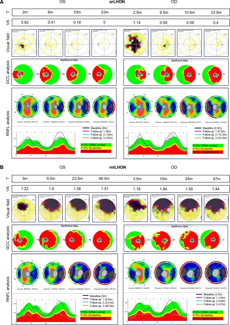
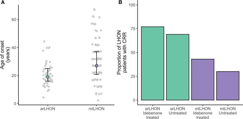
The recent description of biallelic DNAJC30 variants in Leber hereditary optic neuropathy (LHON) and Leigh syndrome challenged the longstanding assumption for LHON to be exclusively maternally inherited and broadened the genetic spectrum of Leigh syndrome, the most frequent paediatric mitochondrial disease. Herein, we characterize 28 so far unreported individuals from 26 families carrying a homozygous DNAJC30 p.Tyr51Cys founder variant, 24 manifesting with LHON, two manifesting with Leigh syndrome, and two remaining asymptomatic. This collection of unreported variant carriers confirms sex-dependent incomplete penetrance of the homozygous variant given a significant male predominance of disease and the report of asymptomatic homozygous variant carriers. The autosomal recessive LHON patients demonstrate an earlier age of disease onset and a higher rate of idebenone-treated and spontaneous recovery of vision in comparison to reported figures for maternally inherited disease. Moreover, the report of two additional patients with childhood- or adult-onset Leigh syndrome further evidences the association of DNAJC30 with Leigh syndrome, previously only reported in a single childhood-onset case.

Keywords: DNAJC30; LHON; Leigh syndrome; mitochondrial disease.


Figures

Figure 1
Figure 1
Ophthalmological investigation of arLHON and mtLHON. Illustrative example of (A) arLHON (DNAJC30, p.Tyr51Cys) and (B) mtLHON (MT-ND4, m.11778G>А) patients at first investigation and subsequent follow-up investigation. Time from symptom onset (T) is indicated in months (m). Visual acuity (VA) was assessed using the logMAR scale and demonstrates visual impairment in arLHON and mtLHON. The arLHON patient demonstrates subsequent complete (OS) or partial (OD) restoration of vision. Visual field was studied by perimetry (Low Vision Center program Octopus 900, Interzeag AG) and demonstrates bilateral central scotomas in arLHON and mtLHON as well as gradual decrease in size and an increase in light sensitivity in arLHON. Analysis of the thickness of the ganglion cell complex (GCC) and the peripapillary layer of the retinal nerve fibre layer (RNFL; RTVue-100 optical coherence tomography, Optovue) demonstrates marked thinning of the GCC and subacute phase swelling of the RNFL, followed by chronic phase thinning of the RNFL in mtLHON that is less pronounced in arLHON. The RNFL thickness graphs display the RNFL thickness values in micrometres in the temporal (T), superior (S), nasal (N) and inferior (I) sectors in the first and subsequent follow-up investigations (visits presented as black, pink, blue and brown curves). OD = ocular dextra (right eye); OS = ocular sinister (left eye); SSI = signal strength index.
Figure 2
Figure 2
Comparison of disease onset and clinically relevant recovery rates of visual acuity in arLHON and mtLHON patients. (A) Reported age of onset for arLHON (n = 53, data unavailable for three patients) and mtLHON (n = 104). (B) Clinically relevant recovery rates for idebenone-treated (n = 30) and untreated (n = 16) arLHON patients (data presented for 46 of 56 arLHON patients with follow-up data available over at least 6 months following onset) and idebenone-treated (n = 184) and untreated (n = 88) mtLHON patients.
Figure 3
Figure 3
MRI brain images from three patients with Leigh syndrome due to DNAJC30 defect. (A) MRI brain images from the first reported female childhood-onset Leigh syndrome patient (Patient 1) taken at 7 years of age demonstrating bilateral signal intensity changes in the putamina and the pedunculi cerebelli (arrows).(B) MRI brain images from the second reported female childhood-onset Leigh syndrome patient (Patient 2) taken at 12 years of age demonstrating bilateral signal intensity changes in the putamina and the heads of caudate nuclei (arrows). The volume of the putamina and caudate heads is decreased bilaterally. (C) MRI brain images from the male adult-onset DNAJC30-associated Leigh syndrome patient (Patient 3) taken at 24 years of age demonstrating bilateral signal intensity changes in the posterior basal ganglia (arrows).

References

    1. Carelli V, Carbonelli M, Irenaeus F, et al. . International consensus statement on the clinical and therapeutic management of Leber hereditary optic neuropathy. J Neuroophthalmol. 2017;37(4):371–381. - PubMed
    1. Newman NJ. Treatment of hereditary optic neuropathies. Nat Rev Neurol. 2012;8(10):545–556. - PubMed
    1. Yu-Wai-Man P, Griffiths PG, Chinnery PF. Mitochondrial optic neuropathies—Disease mechanisms and therapeutic strategies. Prog Retin Eye Res. 2011;30(2):81–114. - PMC - PubMed
    1. Barboni P, Savini G, Valentino ML, et al. . Leber’s hereditary optic neuropathy with childhood onset. Invest Ophthalmol Vis Sci. 2006;47(12):5303–5309. - PubMed
    1. Man PYW, Griffiths PG, Brown DT, Howell N, Turnbull DM, Chinnery PF. The epidemiology of Leber hereditary optic neuropathy in the North East of England. Am J Hum Genet. 2003;72(2):333–339. - PMC - PubMed

Publication types

Substances

Supplementary concepts

LinkOut - more resources