

Accuracy of bone-level assessments following reconstructive surgical treatment of experimental peri-implantitis
- PMID: 35148451
- PMCID: PMC9306925
- DOI: 10.1111/clr.13903
Accuracy of bone-level assessments following reconstructive surgical treatment of experimental peri-implantitis
Abstract
Aims: The purpose of this study was to evaluate the accuracy of bone-level assessments using either cone-beam computed tomography (CBCT), intra-oral peri-apical (PA) radiographs or histology following reconstructive treatment of experimental peri-implantitis.
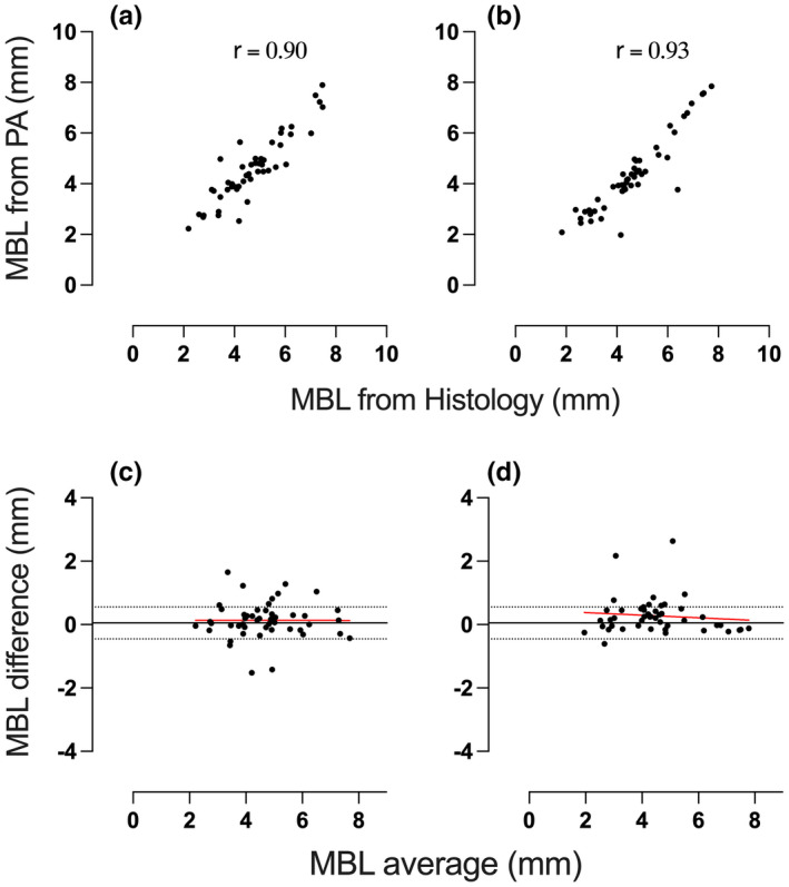
Materials and methods: Six Labrador dogs were used. Experimental peri-implantitis was induced 3 months after implant placement. Surgical treatment of peri-implantitis was performed and peri-implant defects were allocated to one of four treatment categories; no augmentation, bone graft materials with or without a barrier membrane. Six months later, intra-oral PA radiographs and block biopsies from all implants sites were obtained. Marginal bone levels (MBLs) were measured using PA radiographs, CBCT and histology.
Results: Significant correlations of MBL assessments were observed between the three methods. The measurements in PA radiographs consistently resulted in an overestimation of the bone level of about 0.3-0.4 mm. The agreement between the methods was not influenced by the use of bone substitute materials in the management of the osseous defects.
Conclusions: Although MBL assessments obtained from PA radiographs showed an overestimation compared to MBL assessments on corresponding CBCT images and histological sections, PA radiographs can be considered a reliable technique for peri-implant bone-level evaluations following reconstructive surgical therapy of experimental peri-implantitis.
Keywords: cone-beam computed tomography; experimental peri-implantitis; histology; marginal bone level; radiography; reconstructive therapy.
© 2022 The Authors. Clinical Oral Implants Research published by John Wiley & Sons Ltd.
Conflict of interest statement
Dr. Berglundh reports grants from Swedish Research Council (VR), grants from TUA research funding, Gothenburg, Sweden, non‐financial support from Geistlich Biomaterials, Switzerland, non‐financial support from Dentsply Implants IH AB, Sweden, during the conduct of the study. Grants and Personal fees from Dentsply Implants IH AB, Mölndal, Sweden, outside the submitted work. Dr. Abrahamsson reports non‐financial support from Dentsply Sirona, non‐financial support from Geistlich Pharma AG, during the conduct of the study. Grants from Dentsply Sirona, outside the submitted work. Dr. Almohandes, Dr. Lund, Dr. Carcuac and Dr. Petzold have nothing to disclose.
Figures


References
-
- Albouy, J.‐P. , Abrahamsson, I. , Persson, L. G. , & Berglundh, T. (2008). Spontaneous progression of peri‐implantitis at different types of implants. An experimental study in dogs. I: Clinical and radiographic observations. Clinical Oral Implants Research, 19(10), 997–1002. 10.1111/j.1600-0501.2008.01589.x - DOI - PubMed
-
- Bender, P. , Salvi, G. E. , Buser, D. , Sculean, A. , & Bornstein, M. M. (2017). Correlation of three‐dimensional radiologic data with subsequent treatment approach in patients with peri‐implantitis: A retrospective analysis. The International Journal of Periodontics & Restorative Dentistry, 37(4), 481–489. 10.11607/prd.2844 - DOI - PubMed
MeSH terms
Substances
Grants and funding
LinkOut - more resources
Full Text Sources
Medical
Miscellaneous
