. 2022 Jul;101(7):859-869.
doi: 10.1177/00220345221074356. Epub 2022 Feb 11.

Transcriptional Regulation of Jaw Osteoblasts: Development to Pathology

Affiliations

Transcriptional Regulation of Jaw Osteoblasts: Development to Pathology

A Nassif et al. J Dent Res. 2022 Jul.

Abstract

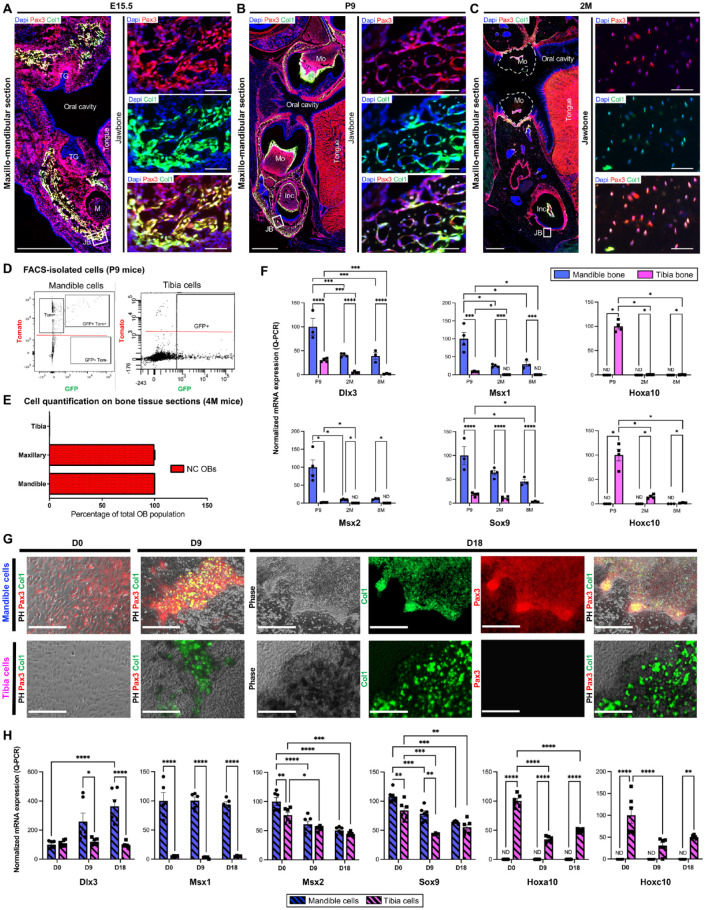
Craniofacial and jaw bones have unique physiological specificities when compared to axial and appendicular bones. However, the molecular profile of the jaw osteoblast (OB) remains incomplete. The present study aimed to decipher the bone site-specific profiles of transcription factors (TFs) expressed in OBs in vivo. Using RNA sequencing analysis, we mapped the transcriptome of confirmed OBs from 2 different skeletal sites: mandible (Md) and tibia (Tb). The OB transcriptome contains 709 TF genes: 608 are similarly expressed in Md-OB and Tb-OB, referred to as "OB-core"; 54 TF genes are upregulated in Md-OB, referred to as "Md-set"; and 18 TF genes are upregulated in Tb-OB, referred to as "Tb-set." Notably, the expression of 29 additional TF genes depends on their RNA transcript variants. TF genes with no previously known role in OBs and bone were identified. Bioinformatics analysis combined with review of genetic disease databases and a comprehensive literature search showed a significant contribution of anatomical origin to the OB signatures. Md-set and Tb-set are enriched with site-specific TF genes associated with development and morphogenesis (neural crest vs. mesoderm), and this developmental imprint persists during growth and homeostasis. Jaw and tibia site-specific OB signatures are associated with craniofacial and appendicular skeletal disorders as well as neurocristopathies, dental disorders, and digit malformations. The present study demonstrates the feasibility of a new method to isolate pure OB populations and map their gene expression signature in the context of OB physiological environment, avoiding in vitro culture and its associated biases. Our results provide insights into the site-specific developmental pathways governing OBs and identify new major OB regulators of bone physiology. We also established the importance of the OB transcriptome as a prognostic tool for human rare bone diseases to explore the hidden pathophysiology of craniofacial malformations, among the most prevalent congenital defects in humans.

Keywords: RNA-seq; bone disease; jawbone; tibia; transcription factor; transcriptome.


Conflict of interest statement

Declaration of Conflicting Interests: The authors declared no potential conflicts of interest with respect to the research, authorship, and/or publication of this article.

Figures

Figure 1.
Figure 1.
Osteoblasts display a bone site-specific transcriptional profile and mesenchymal identity in their physiological environment. (A) Workflow of the RNA sequencing transcriptional study of Md-OBs and Tb-OBs. (B) Total number of TFs commonly expressed (OB-core) or overexpressed by Md-OBs (Md-set) or by Tb-OBs (Tb-set). (C–E) Genes are classified according to their tissue affinities. (C) GO and enrichment analysis of TFs associated with mesenchymal tissues/cells in OB-core, Md-set, and Tb-set (data are presented as –log10 [P value]) pos., positive; neg. negative. (D) RPKM expression heat map of TF genes in Md-set associated with mesenchymal tissues/cells (only RPKM values >1 are shown). RPKM, reads per kilobase of transcript per million mapped reads. *Site-specific genes (all the expressed transcripts of a given gene are increased in Md-OB). (E) Genes known to be involved in different mesenchymal tissues but reported here for the first time as regulators of OBs and bone for the Md-set. (F) Quantitative reverse transcriptase–polymerase chain reaction analysis of in vivo gene expression of Sp6 in tissues isolated from C57BL/6JR mice at E10.5, E14.5, E16.5, E18.5, P1, and P9. Messenger RNA levels are expressed as percentage of TF expression in P9 Md-bone. ND, Non detected, ***p ≤ 0.001. (G–I) Fluorescence imaging of functional OBs (GFP+) and Sp6-expressing cells (pink+) in the BA1 at E10.5 (G) and in the JB at E15.5 (H) and P9 (I) in Col1a1*2,3-GFP mice (scale bars: 200 μm for the main images, 50 μm for the inserts). BA1, first branchial arch; BP, biological process; GO, Gene Ontology; HL, hindlimb; Inc, incisor; JB, jawbone; M, Meckel’s cartilage; Md, mandible; Mo, molar; NT, neural tube; OB, osteoblast; OV, otic vesicle; Tb, tibia; TF, transcription factor; vXX, transcript variant identified by the last 2 numbers of the messenger RNA sequence NM number.
Figure 2.
Figure 2.
The osteoblast transcriptome is characterized by site-specific developmental imprints. (A) GO and enrichment analysis of TF genes associated with developmental processes and morphogenesis in OB-core, Md-set, and Tb-set. (B) Venn diagram of TF genes associated with neural crest (NC) cells in OB-core, Md-set, and Tb-set. (C) RPKM expression heatmap of TF genes in Md-set and Tb-set associated with NC cells (only RPKM values >1 are shown). RPKM, reads per kilobase of transcript per million mapped reads; vXX, transcript variant identified by the last 2 numbers of the mRNA sequence NM number. (D, E) Quantitative reverse transcriptase–polymerase chain reaction analysis of in vivo gene expression of Dlx3 (D) and Sox9 (E) in tissues isolated from C57BL/6JR mice at E10.5, E14.5, E16.5, E18.5, P1, and P9. Messenger RNA levels are expressed as percentage of TF expression in P9 Md-bone. **p ≤ 0.01, ***p ≤ 0.001. (F–H) Fluorescence imaging of functional OBs (GFP+) and Sox9-expressing cells (pink+) in BA1 at E10.5 (F) and JB at E15.5 (G) and P9 (H) in Col1a1*2,3-GFP mice (scale bars: 200 μm for the main images, 50 μm for the inserts). BA1, first branchial arch; BP, biological process; CNC, cranial neural crest; EMT, epithelial to mesenchymal transition; GO, Gene Ontology; HL, hindlimb; Inc, incisor; JB, jawbone; M, Meckel’s cartilage; Md, mandible; Mo, molar; NC, neural crest; NT, neural tube; OB, osteoblast; OV, otic vesicle; Tb, Tibia; TF, transcription factor.
Figure 3.
Figure 3.
Osteoblast embryonic positional identity is maintained in vivo and ex vivo. (A–D) Fluorescence imaging of functional OBs (GFP+) and NC-derived cells (Tomato+) in jawbone at embryonic (E15.5) (A), postnatal (P9) (B), and 2 mo (2M) (C) stages in Pax3-cre::Col1a1*2,3-GFP::Rosatomato mice (scale bars: 500 μm for the main images, 50 μm for the inserts). (D) Double labeling flow cytometry for GFP and Tomato of cells isolated from mandible and tibia of P9 Pax3-cre::Col1a1*2,3-GFP::Rosatomato mice. (E) Quantification (cell counting) of NC-derived cells (tomato+) in mandible, maxillary, and tibia from Pax3-cre::Col1a1*2,3-GFP::Rosatomato mice at 4M. (F) Quantitative reverse transcriptase–polymerase chain reaction (RT-qPCR) analysis of in vivo gene expression of Dlx3, Msx1, Msx2, Sox9, Hoxa10, and Hoxc10 in mandible and tibia bone tissues isolated from C57BL/6JR mice at P9, 2M, and 8 mo (8M). Messenger RNA (mRNA) levels are expressed as percentage of TF expression in P9 Md-bone (for Dlx3, Msx1, Msx2, Sox9) or in P9 Tb-Bone (for Hoxa10 and Hoxc10). (G) Primary cell culture of osteoprogenitors isolated from mandible and tibia of P9 Pax3-cre::Col1a1*2,3-GFP::Rosatomato mice. At confluency, cells were grown in osteogenic medium and observed at day 0 (D0), day 9 (D9), and day 18 (D18) (scale bar: 400 μm). (H) RT-qPCR analysis of ex vivo gene expression of Dlx3, Msx1, Msx2, Sox9, Hoxa10, and Hoxc10 in mandible and tibia osteoprogenitors. mRNA levels are expressed in percentage of TF expression in D0 mandible cells (for Dlx3, Msx1, Msx2, Sox9) or in D0 tibia cells (for Hoxa10 and Hoxc10). ND, Non detected, *p < 0.05, **p ≤ 0.01, ***p ≤ 0.001, ****p ≤ 0.0001. Inc, incisor; JB, jawbone; M, Meckel’s cartilage; Md, mandible; Mo, molar; NC, neural crest; OB, osteoblast; PH, phase; Tb, tibia; TF, transcription factor; TG, tooth germ.
Figure 4.
Figure 4.
Human mutations associated with TF genes of jaw and tibia site-specific osteoblast signatures and their consequences in pathophysiology. (A) RPKM expression heatmap of TF genes in Md-set and Tb-set associated with human genetic disorders (only RPKM values >1 are shown). Rpkm: Reads Per Kilobase of transcript per Million mapped reads, *site-specific genes (all the expressed transcripts of a given gene are commonly expressed in Md-OBs and Tb-OBs), vXX: transcript variant identified by the last 2 numbers of the mRNA sequence NM number. (B, C) OMIM- and Orphanet-based gene-by-gene analysis of genetic disorders and conditions caused by human mutations of Md-set and Tb-set according to tissue type and anatomical region. Pathological growth modification patterns are indicated with yellow arrows. (D) Quantitative reverse transcriptase–polymerase chain reaction analysis of in vivo gene expression of Alx3, Pax3, and Pax9 in mandible and tibia bone tissues isolated from C57BL/6JR mice at postnatal (P9), 2 mo (2M), and 8 mo (8M) stages. Messenger RNA (mRNA) levels are expressed as percentage of TF expression in P9 Md-bone. Md, mandible; OB, osteoblast; Tb, tibia; TF, transcription factor; ND, Non detected, p > 0.5 (ns), *p ≤ 0.05, **p ≤ 0.01, ****p ≤ 0.0001. Abbreviations of human genetic disorders: AI, amelogenesis imperfecta; AML, acute myelogenous leukemia; AMS, ablepharon-macrostomia syndrome; BRMUTD, brain malformations with or without urinary tract defects/1p31p32 microdeletion syndrome; CAKUTHED, congenital anomalies of kidney and urinary tract syndrome with or without hearing loss, abnormal ears, or developmental delay; CDAN, congenital dyserythropoietic anemia (type IV); CDHS, craniofacial-deafness-hand syndrome; CRS, craniosynostosis (types 3 and 5); DRRS, Duane-radial ray syndrome (Okihiro syndrome); FFDD3, focal facial dermal dysplasia 3; FND, frontonasal dysplasia (types 1, 2, and 3); GCPS, Greig cephalopolysyndactyly syndrome; ICF2, immunodeficiency-centromeric instability-facial anomalies syndrome 2; INLU, blood group–Lutheran inhibitor; IVIC, Instituto Venezolano de Investigaciones Cientificas; MDS, myelodysplastic syndrome; OFC, orofacial cleft (type 5); PAP, polydactyly, postaxial (types A1 and B); PFM, parietal foramina 1; PFMCCD, parietal foramina with cleidocranial dysplasia; PHS, Pallister–Hall syndrome; PPD, preaxial postdactyly (type 4); PFM, parietal foramina; RMS, rhabdomyosarcoma 2, alveolar; SAMS, short stature, auditory canal atresia, mandibular hypoplasia, skeletal abnormalities; SHFM, split hand/foot malformation; STHA, selective tooth agenesis (type 3); TDO, trichodontoosseous syndrome; WS, Waardenburg syndrome, type 1.

References

    1. Aghaloo TL, Chaichanasakul T, Bezouglaia O, Kang B, Franco R, Dry SM, Atti E, Tetradis S. 2010. Osteogenic potential of mandibular vs. long-bone marrow stromal cells. J Dent Res. 89(11):1293–1298. - PMC - PubMed
    1. Aïoub M, Lézot F, Molla M, Castaneda B, Robert B, Goubin G, Néfussi JR, Berdal A. 2007. Msx2–/– transgenic mice develop compound amelogenesis imperfecta, dentinogenesis imperfecta and periodental osteopetrosis. Bone. 41(5):851–859. - PubMed
    1. Akintoye SO, Lam T, Shi S, Brahim J, Collins MT, Robey PG. 2006. Skeletal site-specific characterization of orofacial and iliac crest human bone marrow stromal cells in same individuals. Bone. 38(6):758–768. - PubMed
    1. Bertl K, Bertl MH, Heimel P, Burt M, Gahleitner A, Stavropoulos A, Ulm C. 2018. Alveolar bone resorption after primary tooth loss has a negative impact on straightforward implant installation in patients with agenesis of the lower second premolar. Clin Oral Implants Res. 29(2):155–163. - PubMed
    1. Beverdam A, Brouwer A, Reijnen M, Korving J, Meijlink F. 2001. Severe nasal clefting and abnormal embryonic apoptosis in Alx3/Alx4 double mutant mice. Development. 128(20):3975–3986. - PubMed

Publication types

Substances