. 2022 Apr 1;21(4):635-646.
doi: 10.1158/1535-7163.MCT-21-0554.

DS-7300a, a DNA Topoisomerase I Inhibitor, DXd-Based Antibody-Drug Conjugate Targeting B7-H3, Exerts Potent Antitumor Activities in Preclinical Models

Affiliations

DS-7300a, a DNA Topoisomerase I Inhibitor, DXd-Based Antibody-Drug Conjugate Targeting B7-H3, Exerts Potent Antitumor Activities in Preclinical Models

Michiko Yamato et al. Mol Cancer Ther. .

Abstract

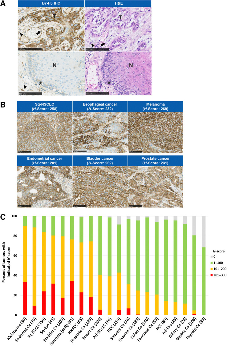
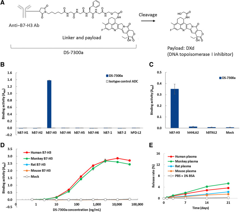
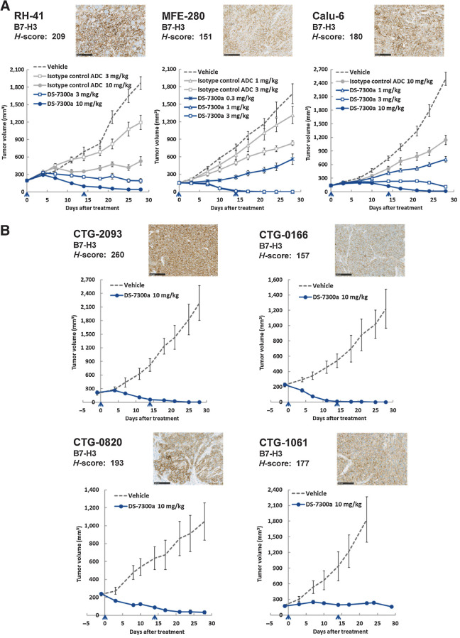
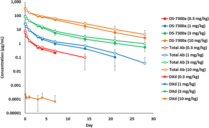
B7-H3 is overexpressed in various solid tumors and has been considered as an attractive target for cancer therapy. Here, we report the development of DS-7300a, a novel B7-H3-targeting antibody-drug conjugate with a potent DNA topoisomerase I inhibitor, and its in vitro profile, pharmacokinetic profiles, safety profiles, and in vivo antitumor activities in nonclinical species. The target specificity and species cross-reactivity of DS-7300a were assessed. Its pharmacologic activities were evaluated in several human cancer cell lines in vitro and xenograft mouse models, including patient-derived xenograft (PDX) mouse models in vivo. Pharmacokinetics was investigated in cynomolgus monkeys. Safety profiles in rats and cynomolgus monkeys were also assessed. DS-7300a specifically bound to B7-H3 and inhibited the growth of B7-H3-expressing cancer cells, but not that of B7-H3-negative cancer cells, in vitro. Additionally, treatment with DS-7300a and DXd induced phosphorylated checkpoint kinase 1, a DNA damage marker, and cleaved PARP, an apoptosis marker, in cancer cells. Moreover, DS-7300a demonstrated potent in vivo antitumor activities in high-B7-H3 tumor xenograft models, including various tumor types of high-B7-H3 PDX models. Furthermore, DS-7300a was stable in circulation with acceptable pharmacokinetic profiles in monkeys, and well tolerated in rats and monkeys. DS-7300a exerted potent antitumor activities against B7-H3-expressing tumors in in vitro and in vivo models, including PDX mouse models, and showed acceptable pharmacokinetic and safety profiles in nonclinical species. Therefore, DS-7300a may be effective in treating patients with B7-H3-expressing solid tumors in a clinical setting.


Figures

Figure 1. IHC analysis of B7-H3 expression on human tumor tissues. A, Representative B7-H3 expression pattern in TMA tissue by B7-H3 IHC and image of hematoxylin and eosin (H&E) staining. Esophageal cancer (top) and normal esophagus tissue adjacent to tumor (bottom). Arrow, vasculature/endothelium; arrowhead, fibrous stromal cells; asterisk, basal cells. Scale bar, 100 μm. B, Representative B7-H3 IHC images of human tumor tissues. Scale bar, 100 μm. C, B7-H3 expression on various human solid tumors. B7-H3 IHC analysis was performed using TMAs. H-score of B7-H3 expression on tumor cells in each sample was calculated. Number in parentheses indicates the number of samples. Ad, adenocarcinoma; ca, cancer; Endomet, endometrial; Eso, esophageal cancer; HCC, hepatocellular carcinoma; HNSCC, head and neck squamous cell carcinoma; N, nontumor epithelium; RCC, renal cell carcinoma; Sq, squamous; T, tumor cells.
Figure 1.
IHC analysis of B7-H3 expression on human tumor tissues. A, Representative B7-H3 expression pattern in TMA tissue by B7-H3 IHC and image of hematoxylin and eosin (H&E) staining. Esophageal cancer (top) and normal esophagus tissue adjacent to tumor (bottom). Arrow, vasculature/endothelium; arrowhead, fibrous stromal cells; asterisk, basal cells. Scale bar, 100 μm. B, Representative B7-H3 IHC images of human tumor tissues. Scale bar, 100 μm. C, B7-H3 expression on various human solid tumors. B7-H3 IHC analysis was performed using TMAs. H-score of B7-H3 expression on tumor cells in each sample was calculated. Number in parentheses indicates the number of samples. Ad, adenocarcinoma; ca, cancer; Endomet, endometrial; Eso, esophageal cancer; HCC, hepatocellular carcinoma; HNSCC, head and neck squamous cell carcinoma; N, nontumor epithelium; RCC, renal cell carcinoma; Sq, squamous; T, tumor cells.
Figure 2. Characteristics of DS-7300a. A, Schematic structure of DS-7300a. B and C, Binding activity of DS-7300a against human B7 family proteins. Recombinant proteins of human B7-H1, B7-H2, B7-H3 (4Ig), B7-H4, B7-H5, B7-H6, B7–1, B7–2, and PD-L2 were incubated with DS-7300a or isotype control ADC and binding activities were measured by ELISA. CHO-K1 cells expressing human B7-H3 (4Ig), HHLA2, and BTNL2 were incubated with DS-7300a and binding activities were measured by cell-based ELISA. Each value represents the mean and SD (N = 3). D, Species cross-reactivity of DS-7300a. CHO-K1 cells expressing human (4Ig), cynomolgus monkey (4Ig), rat, and mouse B7-H3 were incubated with DS-7300a and binding activities were evaluated by cell-based ELISA. Each value represents the mean and SD (N = 3). E, In vitro stability of DS-7300a in plasma. The release rate of DXd from 100 μg/mL DS-7300a in human, monkey, rat, and mouse plasma was calculated using the mean concentration of the released DXd (N = 3).
Figure 2.
Characteristics of DS-7300a. A, Schematic structure of DS-7300a. B and C, Binding activity of DS-7300a against human B7 family proteins. Recombinant proteins of human B7-H1, B7-H2, B7-H3 (4Ig), B7-H4, B7-H5, B7-H6, B7–1, B7–2, and PD-L2 were incubated with DS-7300a or isotype control ADC and binding activities were measured by ELISA. CHO-K1 cells expressing human B7-H3 (4Ig), HHLA2, and BTNL2 were incubated with DS-7300a and binding activities were measured by cell-based ELISA. Each value represents the mean and SD (N = 3). D, Species cross-reactivity of DS-7300a. CHO-K1 cells expressing human (4Ig), cynomolgus monkey (4Ig), rat, and mouse B7-H3 were incubated with DS-7300a and binding activities were evaluated by cell-based ELISA. Each value represents the mean and SD (N = 3). E,In vitro stability of DS-7300a in plasma. The release rate of DXd from 100 μg/mL DS-7300a in human, monkey, rat, and mouse plasma was calculated using the mean concentration of the released DXd (N = 3).
Figure 3. In vitro cytotoxic effects of DS-7300a. A and B, B7-H3 expression on human cancer cells and in vitro cell growth inhibition by DS-7300a. RH-41 (rhabdomyosarcoma), MFE-280 (endometrial adenocarcinoma), and CCRF-CEM (acute lymphocytic leukemia) cells were stained with PE-conjugated isotype control (mIgG2b, gray open histogram) or antihuman B7-H3 antibody (gray closed histogram) and then analyzed by flow cytometry. Cells were cultured with DS-7300a, parental anti–B7-H3 Ab, and isotype control ADC for 6 days, and then cell viability was examined. Each value represents the mean and SD (N = 3). C, DNA damage and apoptosis induced by DS-7300a. RH-41 cells were treated with 10 μg/mL DS-7300a, parental anti–B7-H3 Ab, isotype control ADC, or 10 nmol/L DXd for 72 hours. Then, the cells were harvested and analyzed by Western blotting for phosphorylated Chk1 (pChk1) as a DNA damage marker and for cleaved PARP as an apoptosis marker.
Figure 3.
In vitro cytotoxic effects of DS-7300a. A and B, B7-H3 expression on human cancer cells and in vitro cell growth inhibition by DS-7300a. RH-41 (rhabdomyosarcoma), MFE-280 (endometrial adenocarcinoma), and CCRF-CEM (acute lymphocytic leukemia) cells were stained with PE-conjugated isotype control (mIgG2b, gray open histogram) or antihuman B7-H3 antibody (gray closed histogram) and then analyzed by flow cytometry. Cells were cultured with DS-7300a, parental anti–B7-H3 Ab, and isotype control ADC for 6 days, and then cell viability was examined. Each value represents the mean and SD (N = 3). C, DNA damage and apoptosis induced by DS-7300a. RH-41 cells were treated with 10 μg/mL DS-7300a, parental anti–B7-H3 Ab, isotype control ADC, or 10 nmol/L DXd for 72 hours. Then, the cells were harvested and analyzed by Western blotting for phosphorylated Chk1 (pChk1) as a DNA damage marker and for cleaved PARP as an apoptosis marker.
Figure 4. Antitumor activities of DS-7300a in xenograft mouse models. Representative B7-H3 IHC images of tumors and H-scores were also shown for each model. Scale bar, 100 μm. The indicated H-score represents the mean of H-scores in three tumor samples. The arrowhead indicates the time point of treatment. Each value represents the mean and SE of tumor volume. A, Antitumor activities of DS-7300a in cell line–derived xenograft models. Mice inoculated with human cancer cells, RH-41, MFE-280, or Calu-6 (NSCLC) were treated with DS-7300a, vehicle control, or isotype control ADC intravenously on days 0 and 14 (N = 6 mice/group). B, Antitumor activities of DS-7300a in various tumor types of PDX models. PDX mice were treated with 10 mg/kg DS-7300a or vehicle control intravenously on days 0 and 14 (N = 5 mice/group). CTG-2093, SCLC PDX; CTG-0166, NSCLC PDX; CTG-0820, head and neck cancer PDX; CTG-1061, bladder cancer PDX.
Figure 4.
Antitumor activities of DS-7300a in xenograft mouse models. Representative B7-H3 IHC images of tumors and H-scores were also shown for each model. Scale bar, 100 μm. The indicated H-score represents the mean of H-scores in three tumor samples. The arrowhead indicates the time point of treatment. Each value represents the mean and SE of tumor volume. A, Antitumor activities of DS-7300a in cell line–derived xenograft models. Mice inoculated with human cancer cells, RH-41, MFE-280, or Calu-6 (NSCLC) were treated with DS-7300a, vehicle control, or isotype control ADC intravenously on days 0 and 14 (N = 6 mice/group). B, Antitumor activities of DS-7300a in various tumor types of PDX models. PDX mice were treated with 10 mg/kg DS-7300a or vehicle control intravenously on days 0 and 14 (N = 5 mice/group). CTG-2093, SCLC PDX; CTG-0166, NSCLC PDX; CTG-0820, head and neck cancer PDX; CTG-1061, bladder cancer PDX.
Figure 5. Pharmacokinetics of DS-7300a in monkeys. DS-7300a was administered once intravenously at 0.3, 1, 3, and 10 mg/kg to cynomolgus monkeys. Plasma concentrations of DS-7300a, total Ab, and DXd were determined. Each value represents the mean and SD (N = 3).
Figure 5.
Pharmacokinetics of DS-7300a in monkeys. DS-7300a was administered once intravenously at 0.3, 1, 3, and 10 mg/kg to cynomolgus monkeys. Plasma concentrations of DS-7300a, total Ab, and DXd were determined. Each value represents the mean and SD (N = 3).

References

    1. Chapoval AI, Ni J, Lau JS, Wilcox RA, Flies DB, Liu D, et al. . B7-H3: a costimulatory molecule for T cell activation and IFN-γ production. Nat Immunol 2001;2:269–74. - PubMed
    1. Schildberg FA, Klein SR, Freeman GJ, Sharpe AH. Coinhibitory pathways in the B7-CD28 ligand-receptor family. Immunity 2016;44:955–72. - PMC - PubMed
    1. Sun M, Richards S, Prasad DVR, Mai XM, Rudensky A, Dong C. Characterization of mouse and human B7-H3 genes. J Immunol 2002;168:6294–7. - PubMed
    1. Ling V, Wu PW, Spaulding V, Kieleczawa J, Luxenberg D, Carreno BM, et al. . Duplication of primate and rodent B7-H3 immunoglobulin V- and C-like domains: divergent history of functional redundancy and exon loss. Genomics 2003;82:365–77. - PubMed
    1. Steinberger P, Majdic O, Derdak SV, Pfistershammer K, Kirchberger S, Klauser C, et al. . Molecular characterization of human 4Ig-B7-H3, a member of the B7 family with four Ig-like domains. J Immunol 2004;172:2352–9. - PubMed