. 2022 Mar 1;119(9):e2117198119.
doi: 10.1073/pnas.2117198119.

Targeting stem-loop 1 of the SARS-CoV-2 5' UTR to suppress viral translation and Nsp1 evasion

Affiliations

Targeting stem-loop 1 of the SARS-CoV-2 5' UTR to suppress viral translation and Nsp1 evasion

Setu M Vora et al. Proc Natl Acad Sci U S A. .

Abstract

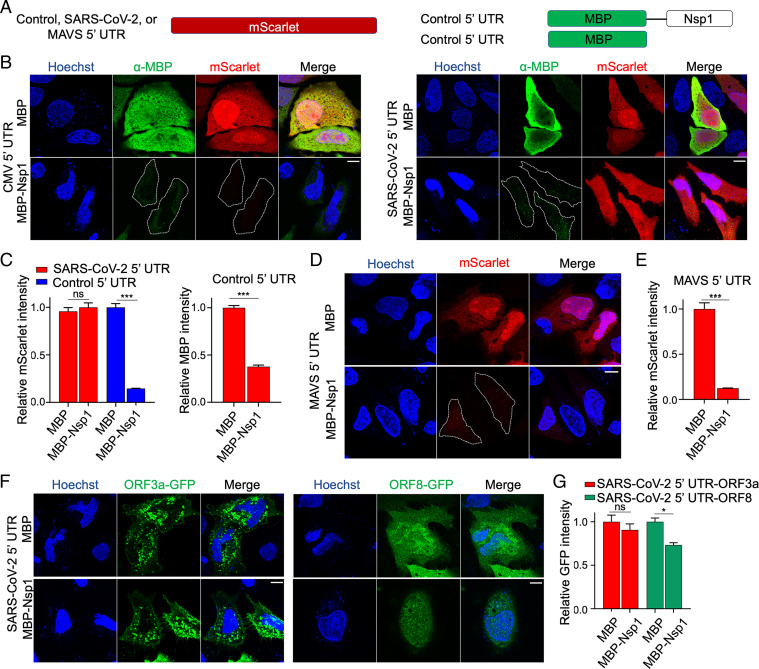
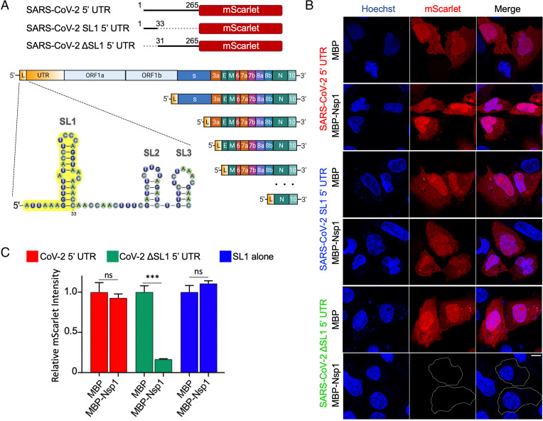
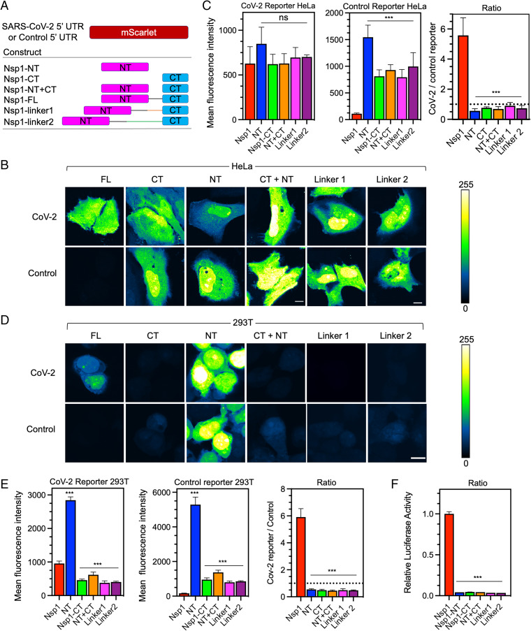
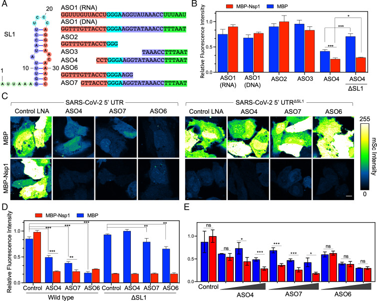
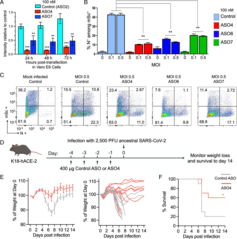
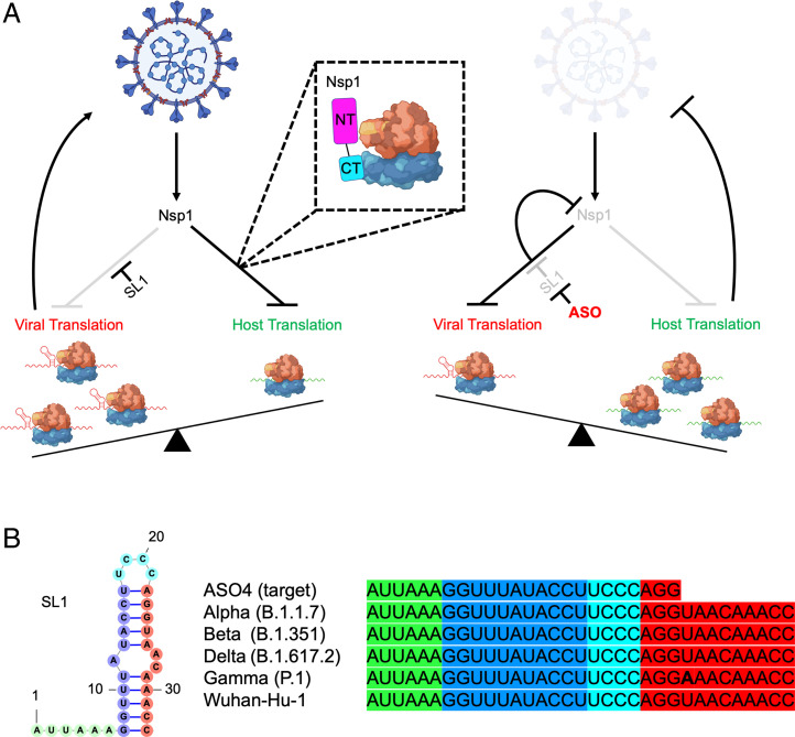
SARS-CoV-2 is a highly pathogenic virus that evades antiviral immunity by interfering with host protein synthesis, mRNA stability, and protein trafficking. The SARS-CoV-2 nonstructural protein 1 (Nsp1) uses its C-terminal domain to block the messenger RNA (mRNA) entry channel of the 40S ribosome to inhibit host protein synthesis. However, how SARS-CoV-2 circumvents Nsp1-mediated suppression for viral protein synthesis and if the mechanism can be targeted therapeutically remain unclear. Here, we show that N- and C-terminal domains of Nsp1 coordinate to drive a tuned ratio of viral to host translation, likely to maintain a certain level of host fitness while maximizing replication. We reveal that the stem-loop 1 (SL1) region of the SARS-CoV-2 5' untranslated region (5' UTR) is necessary and sufficient to evade Nsp1-mediated translational suppression. Targeting SL1 with locked nucleic acid antisense oligonucleotides inhibits viral translation and makes SARS-CoV-2 5' UTR vulnerable to Nsp1 suppression, hindering viral replication in vitro at a nanomolar concentration, as well as providing protection against SARS-CoV-2-induced lethality in transgenic mice expressing human ACE2. Thus, SL1 allows Nsp1 to switch infected cells from host to SARS-CoV-2 translation, presenting a therapeutic target against COVID-19 that is conserved among immune-evasive variants. This unique strategy of unleashing a virus' own virulence mechanism against itself could force a critical trade-off between drug resistance and pathogenicity.

Keywords: SARS-CoV-2; therapeutic; translation.


Conflict of interest statement

The authors declare no competing interest.

Figures

Fig. 1.
Fig. 1.
SARS-CoV-2 5′ UTR bypasses Nsp1-mediated inhibition of translation. (A) Schematic of translational reporters. The 5′ UTR sequences from control, MAVS, or SARS-CoV-2 were placed upstream of the mScarlet reporter (Left). MBP or MBP-Nsp1 (Right) were both downstream of the control 5′ UTR and were cotransfected along with each reporter plasmid. A CMV promoter was used to drive expression in all constructs. (B) Representative images of HeLa cells cotransfected with control 5′ UTR reporter or SARS-CoV-2 5′ UTR reporter and either MBP alone or MBP-Nsp1 and visualized for DNA by Hoechst (blue), MBP by indirect immunofluorescence (green), and mScarlet by in situ fluorescence (red). Successfully transfected cells difficult to visualize due to low intensity are outlined here and in other figures. (C) Quantification of relative mScarlet intensity of data corresponding to B. (D) Representative images of HeLa cells transfected with MAVs 5′ UTR reporter (red). (E) Quantification of relative mScarlet intensity in D. (F) HeLa cells transfected with either ORF3a-GFP (Left) or ORF8-GFP (Right) downstream of SARS-CoV-2 5′ UTR. (G) Quantification of relative GFP intensity in F. Error bars correspond to SEM except where otherwise noted. (Scale bars, 10 μm.) *P < 0.05, **P < 0.01, and ***P < 0.001, Student t test.
Fig. 2.
Fig. 2.
The SL1 stem-loop of the 5′ UTR is necessary and sufficient for evasion of Nsp1-mediated translation suppression. (A) Schematic representation of 5′ UTR, SL1 5′ UTR, and ΔSL1 5′ UTR placed upstream of mScarlet (Upper), and of SARS-CoV-2 leader sequence containing SL1 (yellow) along with its incorporation into subgenomic RNAs (Lower). (B) Representative images of HeLa cells cotransfected with SARS-CoV-2 5′ UTR reporter and either MBP-alone or MBP-Nsp1, and visualized for DNA by Hoechst (blue) and mScarlet by in situ fluorescence (red). (C) Quantification of relative mScarlet intensity of data corresponding to B. (Scale bars, 10 μm.) *P < 0.05, **P < 0.01, and ***P < 0.001, Student t test.
Fig. 3.
Fig. 3.
Nsp1 NT and CT cooperate to drive viral translation selectivity. (A) Schematic of coexpression system with CoV-2 or control reporter along with various fragments of Nsp1 (FL, NT, CT, NT+CT) or extended linker mutants (linker1, linker2). (B) mScarlet fluorescence intensity in HeLa cells cotransfected with either the CoV-2 (Upper) or control reporter (Lower) along with various mutants of Nsp1. Intensity values are fals- colored according to a scale (Right). (Scale bar, 10 μ.) (C) Quantification of fluorescent intensity in B of CoV-2 (Left) or control reporters (Center) and the ratio of CoV-2/Control (Right) with different Nsp1 mutants. (Dashed line marks ratio of 1). (D) mScarlet fluorescence intensity in 293T cells as in B. (E) Quantification of D. (F) Relative luciferase activity in 293T cells assay cotransfected with CoV-2 firefly luciferase and control Renilla luciferase reporters along with various Nsp1 mutants. The ratio of firefly/Renilla luciferase was normalized and plotted in three replicates. Error bars represent SD. (Scale bars, 10 μm.) *P < 0.05, **P < 0.01, and ***P < 0.001, Student t test.
Fig. 4.
Fig. 4.
ASOs targeting SL1 renders the SARS-CoV-2 5′ UTR susceptible to Nsp1-mediated shutdown. (A) Schematic of SL1 region and various ASOs (which are all LNA mixmers unless otherwise noted). Complementary sequences between ASOs and SL1 are matched by color. (B) Initial screen of ASO activity. Each ASO was transfected at 50 nM with CoV-2 reporter along with either MBP-alone or MBP-Nsp1. Bar on far right indicates cotransfection with a reporter lacking SL1 as a control. (C) Images of HeLa cells transfected with 50 nM ASO4, -7, -6, or a control ASO along with CoV-2 or ΔSL1 reporter and either MBP or MBP-Nsp1. Intensity values are false-colored according to a scale (Right). (D) Quantification of C. (E) Dose–response assay of each ASO. Cells were transfected with CoV-2 reporter and MBP or MBP-Nsp1 as above. ASOs were transfected at either 25, 50, or 100 nM. (Scale bars 10 μm.) *P < 0.05, **P < 0.01, and ***P < 0.001, Student t test.
Fig. 5.
Fig. 5.
ASOs targeting SL1 produce stable loss of function to inhibit SARS-CoV-2 replication in vitro and ASO4 provides significant protection against SARS-CoV-2–induced lethality in K18-hACE2 mice. (A) Various ASOs along with CoV-2 reporter and MBP-Nsp1 were transiently transfected into Vero E6 cells and reporter intensity was measured daily over the course of 72 h, shown for each ASO (control, ASO4, and ASO7) at each time point. Since expression from transfected plasmids naturally changes over time, each datapoint was normalized to intensity of a parallel control where no ASO was included. (B) Percent of successfully transfected cells (marked by mScarlet [mSc] positivity) that were N+ by ASO treatment (color coded according to the legend on the right) at various MOIs. Error bars represent SD. (A and B) *P < 0.05, **P < 0.01, and ***P < 0.001, Student t test. (C) Nucleocapsid intensity plotted against mSc obtained by flow cytometry for each treatment (infected at MOI 0.5). Quadrants demarcate mSc+, N+ cells (top right quadrant), and the corresponding percentage of cells is listed in each corner. (D) Schematic for mouse infection experiment. K18-hACE-2 mice were treated with daily intranasally administered control ASO or ASO4 for 4 d following infection with 2,500 PFU of SARS-CoV-2 and monitored for weight loss and survival for 14 d. (E) Average percent weight loss over time for control ASO (gray) or ASO4 (red) after infection (Left) and individual weight loss trajectories (Right). (F) Survival curves over time for control ASO (gray) or ASO4 (red) after infection. n = 10 mice for each treatment.
Fig. 6.
Fig. 6.
Model for Nsp1-driven viral translation selectivity and its disruption via ASO targeting of the highly conserved SL1 region. (A) Nsp1 shuts down host translation, mainly by blocking the mRNA entry channel of the 40S ribosome which ultimately results in host mRNA degradation. The SL1 region in SARS-CoV-2 5′ UTR allows evasion of translational suppression, leading to selective viral translation. Targeting SL1 via ASO makes SARS-CoV-2 5′ UTR vulnerable to Nsp1-mediated translation suppression, resulting in loss of translation of Nsp1 itself and restoration of host translation, allowing antiviral defense to more effectively halt viral replication. (B) Alignment of the ASO4 target sequence with SL1 sequences from SARS-CoV-2 variants of concern showing complete conservation of the sequence targeted by ASO4.

Update of

References

    1. Chan J. F., et al. , Genomic characterization of the 2019 novel human-pathogenic coronavirus isolated from a patient with atypical pneumonia after visiting Wuhan. Emerg. Microbes Infect. 9, 221–236 (2020). - PMC - PubMed
    1. Chan J. F., et al. , A familial cluster of pneumonia associated with the 2019 novel coronavirus indicating person-to-person transmission: A study of a family cluster. Lancet 395, 514–523 (2020). - PMC - PubMed
    1. Wu F., et al. , A new coronavirus associated with human respiratory disease in China. Nature 579, 265–269 (2020). - PMC - PubMed
    1. Zhu N., et al. ; China Novel Coronavirus Investigating and Research Team, A novel coronavirus from patients with pneumonia in China, 2019. N. Engl. J. Med. 382, 727–733 (2020). - PMC - PubMed
    1. Lu R., et al. , Genomic characterisation and epidemiology of 2019 novel coronavirus: Implications for virus origins and receptor binding. Lancet 395, 565–574 (2020). - PMC - PubMed

Publication types

MeSH terms

Substances