. 2022 Feb 15;119(7):e2119678119.
doi: 10.1073/pnas.2119678119.

Reversible lysine fatty acylation of an anchoring protein mediates adipocyte adrenergic signaling

Affiliations

Reversible lysine fatty acylation of an anchoring protein mediates adipocyte adrenergic signaling

Rushita A Bagchi et al. Proc Natl Acad Sci U S A. .

Abstract

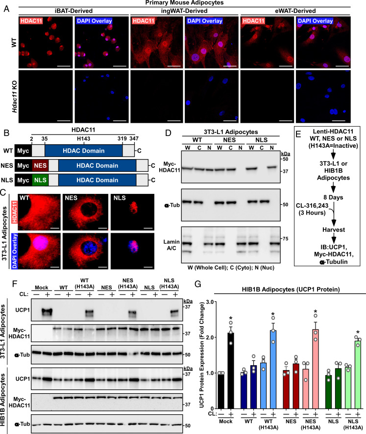
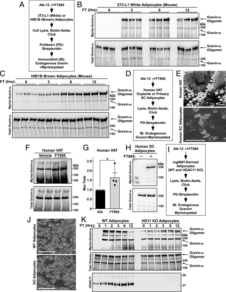
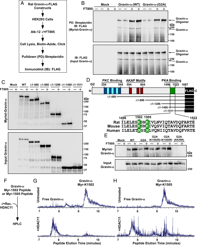
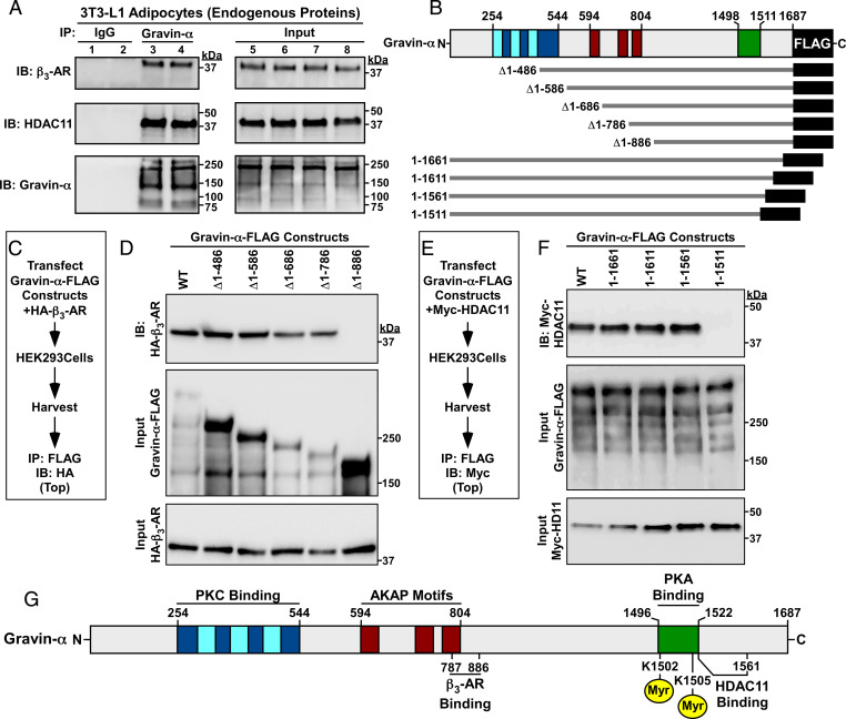
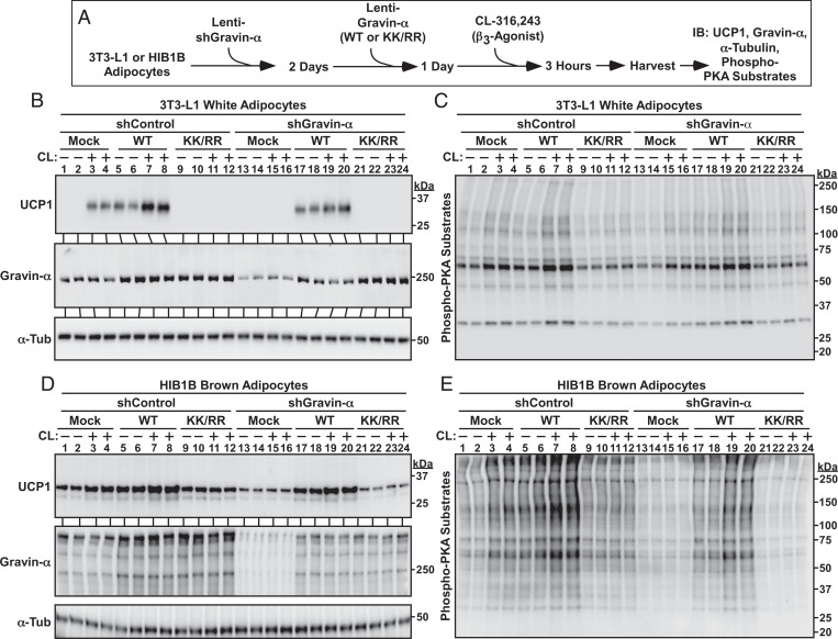
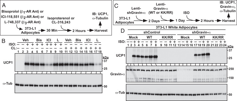
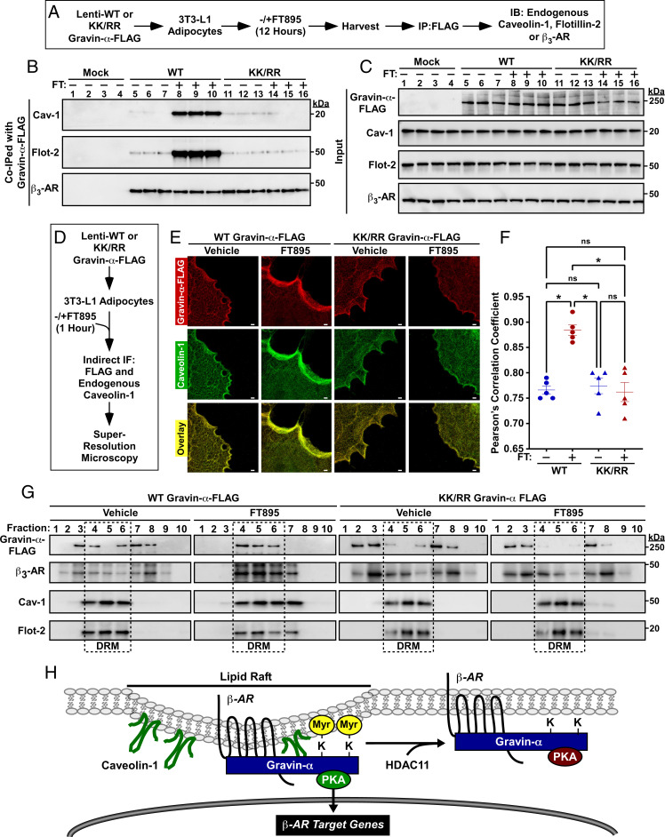
N-myristoylation on glycine is an irreversible modification that has long been recognized to govern protein localization and function. In contrast, the biological roles of lysine myristoylation remain ill-defined. We demonstrate that the cytoplasmic scaffolding protein, gravin-α/A kinase-anchoring protein 12, is myristoylated on two lysine residues embedded in its carboxyl-terminal protein kinase A (PKA) binding domain. Histone deacetylase 11 (HDAC11) docks to an adjacent region of gravin-α and demyristoylates these sites. In brown and white adipocytes, lysine myristoylation of gravin-α is required for signaling via β2- and β3-adrenergic receptors (β-ARs), which are G protein-coupled receptors (GPCRs). Lysine myristoylation of gravin-α drives β-ARs to lipid raft membrane microdomains, which results in PKA activation and downstream signaling that culminates in protective thermogenic gene expression. These findings define reversible lysine myristoylation as a mechanism for controlling GPCR signaling and highlight the potential of inhibiting HDAC11 to manipulate adipocyte phenotypes for therapeutic purposes.

Keywords: HDAC11; adrenergic receptor; lysine myristoylation; signal transduction.


Conflict of interest statement

Competing interest statement: T.A.M. is on the scientific advisory boards of Artemes Bio, Inc., and Eikonizo Therapeutics, received funding from Italfarmaco for an unrelated project, and has a subcontract from Eikonizo Therapeutics related to a Small Business Innovation Research grant from the NIH (HL154959). H.L. is a founder and consultant for Sedec Therapeutics.

Figures

Fig. 1.
Fig. 1.
Nuclear and cytoplasmic HDAC11 are capable of blocking β3-AR–mediated UCP1 induction. (A) Indirect immunofluorescence of endogenous HDAC11 in primary mouse adipocytes derived from adipose depots of WT or Hdac11 KO mice. Nuclei were stained with DAPI. (Scale bar, 50 µm.) (B) Schematic representation of Myc-tagged human HDAC11 expression constructs. Conversion of histidine-143 to alanine (H143A) renders HDAC11 catalytically inactive. (C) Indirect immunofluorescence of Myc-tagged HDAC11 in 3T3-L1 adipocytes. (Scale bar, 10 µm.) (D) Immunoblots confirming expression of Myc-tagged HDAC11 in the predicted subcellular fractions of 3T3-L1 adipocytes. α-Tubulin and lamin A/C served as controls for the purity of cytoplasmic and nuclear fractions, respectively. (E) Schematic depiction of the β3-AR signaling experiment. (F) Immunoblot analysis of 3T3-L1 white adipocyte and HIB1B brown adipocyte homogenates. (G) Quantification of UCP1 protein in three independent experiments with HIB1B cells. Each N = an independent plate of cells. Data are presented as mean + SEM; *P ≤ 0.05 versus unstimulated controls in each condition. Please visit Figshare for a higher-resolution version (10.6084/m9.figshare.19082813).
Fig. 2.
Fig. 2.
Gravin-α/AKAP12 myristoylation is induced upon HDAC11 inhibition. (A) Schematic depiction of the click chemistry experiment employed to assess gravin-α myristoylation in 3T3-L1 and HIB1B adipocytes. Cells were treated with 10 μM FT895 for the indicated times. (B) Immunoblot analysis of 3T3-LI adipocyte homogenates. FT, FT895; Hrs, hours; Myrist, myristoylated. (C) Immunoblot analysis of HIB1B adipocyte homogenates. (D) Schematic depiction of the click chemistry experiment to assess gravin-α myristoylation in human adipose tissue explants and purified adipocytes. Explants and cells were treated with 100 μM and 10 μM FT895 for 12 h, respectively. SC, subcutaneous. (E, Top) Representative whole mount human visceral adipose tissue (VAT) confocal microscopy image with BODIPY 493/503 (lipid droplet) staining; 20× objective lens. (Scale bar, 100 µm.) (Lower) Representative brightfield microscopy image of differentiated human SC stromal vascular fraction (SVF)-derived adipocytes; 10× objective lens. (Scale bar, 400 µm.) (F) Immunoblot analysis of VAT homogenates. (G) Densitometry quantification of myristoylated gravin-α relative to total gravin-α protein in human VAT explants. Data are represented as means +SEM. n = 4 per condition; *P < 0.05 compared to untreated. Veh, vehicle. (H) Immunoblot analysis of human SC adipocyte homogenates. (I) Schematic depiction of the click chemistry experiment to assess gravin-α myristoylation in cultured adipocytes derived from WT and Hdac11 KO ingWAT. Cells were treated with 10 μM FT895 for the indicated times. (J) Brightfield microscopy images of differentiated adipocytes derived from WT and Hdac11 KO ingWAT. (Scale bar, 400 µm.) (K) Immunoblot analysis of homogenates from adipocytes treated for the indicated times with 10 μM FT895. Please visit Figshare for a higher-resolution version (10.6084/m9.figshare.19082813).
Fig. 3.
Fig. 3.
HDAC11 demyristoylates two lysines in the PKA binding domain of gravin-α. (A) Schematic depiction of the experiment to assess myristoylation of ectopically expressed gravin-α constructs. (B) Immunoblot analysis of homogenates of HEK293 cells transfected with the indicated FLAG-tagged gravin-α constructs and treated with vehicle control (−) or 10 μM FT895 for 12 h. Myrist, myristoylated. (C) Immunoblot analysis of homogenates of HEK293 cells transfected with the indicated constructs for N-terminally truncated, FLAG-tagged gravin-α and treated with vehicle control (−) or 10 μM FT895 for 12 h. (D) Schematic representation of gravin-α, gravin-α constructs, and the conserved lysine myristoylation sites embedded in the PKA binding domain of gravin-α. (E) Immunoblot analysis of homogenates of HEK293 cells transfected with constructs for gravin-α harboring the indicated amino acid substitutions and treated with vehicle control (−) or 10 μM FT895 for 12 h. (F) Schematic depiction of the experiment to address whether HDAC11 is capable of directly demyristoylating lysine-1502 and lysine-1505 of gravin-α. (G) LC-MS traces of myristoyl-lysine-1502 peptide and free peptide of gravin-α before and after treatment with recombinant HDAC11 for 1.5 h at 37 °C. (H) LC-MS traces of myristoyl-lysine-1505 peptide and free peptide of gravin-α before and after treatment with recombinant HDAC11 for 1.5 h at 37 °C. For G and H, peaks were searched using Xcaliber software. Myr, myristoyl. Peaks were searched using Xcaliber software. Please visit Figshare for a higher-resolution version (10.6084/m9.figshare.19082813).
Fig. 4.
Fig. 4.
Discrete regions of gravin-α mediate associations with the β3-AR and HDAC11. (A) Co-IP of endogenous gravin-α with endogenous β3-AR and HDAC11 in homogenates from 3T3-L1 adipocytes. IP, immunoprecipitation; IB, immunoblotting. (B) Schematic representation of the N- and carboxyl-terminal truncation constructs of FLAG-tagged rat gravin-α that were employed for co-IP studies. (C) Schematic depiction of the experiment to map the β3-AR binding domain on gravin-α. (D) Immunoblot analysis of ectopically expressed β3-AR coimmunoprecipitating with the indicated gravin-α constructs in HEK293 cell homogenates. (E) Schematic depiction of the experiment to map the HDAC11 binding domain on gravin-α. (F) Immunoblot analysis of ectopically expressed HDAC11 coimmunoprecipitating with the indicated gravin-α constructs in HEK293 cell homogenates. (G) Schematic representation of gravin-α incorporating the β3-AR and HDAC11 binding domains. Please visit Figshare for a higher-resolution version (10.6084/m9.figshare.19082813).
Fig. 5.
Fig. 5.
Lysine myristoylation of gravin-α is required for β3-AR–mediated UCP1 induction. (A) Schematic depiction of the experiment to determine if gravin-α and its myristoylation are required for β3-AR signaling in adipocytes. (B) Immunoblot analysis of UCP1 induction and gravin-α knockdown in 3T3-L1 adipocytes. α-Tubulin (α-Tub) served as a loading control. (C) Immunoblot analysis of 3T3-L1 homogenates using an anti-phospho-PKA substrates antibody. (D) Immunoblot analysis of UCP1 induction and gravin-α knockdown in HIB1B adipocytes. α-Tub served as a loading control. (E) Immunoblot analysis of HIB1B homogenates using an anti-phospho-PKA substrates antibody. Please visit Figshare for a higher-resolution version (10.6084/m9.figshare.19082813).
Fig. 6.
Fig. 6.
Lysine myristoylation of gravin-α is required for β2-AR–mediated UCP1 induction. (A) Schematic depiction of the experiment to address the roles of the three different β-AR isoforms in the control of UCP1 induction. Cells were pretreated with 10 μM bisoprolol, 1 μM ICI-118,551, or 10 μM L-748,337, followed by addition of 1 μM isoproterenol (ISO) or 1 μM CL-316,243. (B) Immunoblot analysis of homogenates from 3T3-L1 adipocytes showing that ISO drives UCP1 induction via β2-AR stimulation, while CL induces UCP1 by promoting β3-AR signaling. Veh, vehicle; Bis, Bisoprolol; ICI, ICI-118,551; L, L-748,337. (C) Schematic depiction of the experiment to determine if gravin-α and its myristoylation are required for β2-AR signaling in adipocytes. (D) Immunoblot analysis of UCP1 induction and gravin-α knockdown in 3T3-L1 adipocytes. α-Tubulin (α-Tub) served as a loading control. Please visit Figshare for a higher-resolution version (10.6084/m9.figshare.19082813).
Fig. 7.
Fig. 7.
Lysine myristoylation of gravin-α targets β3-ARs to caveolin-rich lipid rafts. (A) Schematic depiction of the experiment to address whether lysine myristoylation of gravin-α regulates association with lipid raft proteins, Cav-1 and flotillin-2, and the β3-AR. Cells were treated with 10 μM FT895 or vehicle control. (B) Immunoblot analysis of proteins that coimmunoprecipitated with FLAG-tagged gravin-α WT or KK/RR in 3T3-L1 homogenates. FT, FT895. (C) Immunoblot analysis of input levels of the indicated proteins prior to immunoprecipitation. (D) Schematic depiction of the experiment to determine if HDAC11-reversible myristoylation alters the subcellular localization of gravin-α and/or Cav-1. (E) STED microscopy of FLAG-tagged gravin-α (red) and endogenous Cav-1 (green) in 3T3-L1 adipocytes treated with 10 μM FT895 or vehicle control for 1 h. (Scale bar, 1,000 nm.) KK/RR, myristoylation-deficient. (F) Statistical analysis of the colocalization of ectopic gravin-α with endogenous Cav-1 using Pearson’s correlation coefficient. Each dot represents one cell; center lines represent mean values. FT, FT895. *P < 0.05. ns = not significant. (G) Immunoblot analysis showing ectopic gravin-α and endogenous β3-AR, Cav-1, and Flot-2 in fractions 1 through 10 from detergent-resistant membrane isolation in 3T3L1 adipocytes treated with 10 μM FT895 or vehicle control for 12 h. (H) A model for the regulation of β-AR signaling in adipocytes by reversible lysine myrisotylation. Myristoylation of gravin-α on two lysines in its PKA binding domain targets gravin-α:β-AR:PKA complexes to Cav-1–rich lipid rafts, resulting in downstream signaling and induction of β-AR target genes, including that encoding UCP1. HDAC11-mediated demyristoylation of gravin-α prevents the complexes from localizing to the lipid rafts and thus squelches β-AR signaling. Please visit Figshare for a higher-resolution version (10.6084/m9.figshare.19082813).

Similar articles

Cited by

References

    1. Kosciuk T., Lin H., N-myristoyltransferase as a glycine and lysine myristoyltransferase in cancer, immunity, and infections. ACS Chem. Biol. 15, 1747–1758 (2020). - PMC - PubMed
    1. Jiang H., et al. , Protein lipidation: Occurrence, mechanisms, biological functions, and enabling technologies. Chem. Rev. 118, 919–988 (2018). - PMC - PubMed
    1. Resh M. D., Myristylation and palmitylation of Src family members: The fats of the matter. Cell 76, 411–413 (1994). - PubMed
    1. Dian C., et al. , High-resolution snapshots of human N-myristoyltransferase in action illuminate a mechanism promoting N-terminal Lys and Gly myristoylation. Nat. Commun. 11, 1132 (2020). - PMC - PubMed
    1. Kosciuk T., et al. , NMT1 and NMT2 are lysine myristoyltransferases regulating the ARF6 GTPase cycle. Nat. Commun. 11, 1067 (2020). - PMC - PubMed

LinkOut - more resources