. 2022 Feb 11;13(1):841.
doi: 10.1038/s41467-022-28387-5.

Deep learning-based high-accuracy quantitation for lumbar intervertebral disc degeneration from MRI

Affiliations

Deep learning-based high-accuracy quantitation for lumbar intervertebral disc degeneration from MRI

Hua-Dong Zheng et al. Nat Commun. .

Abstract

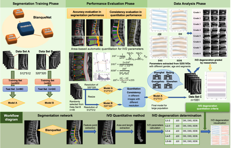
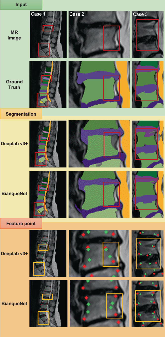
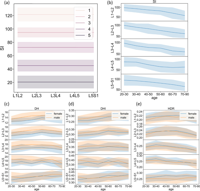
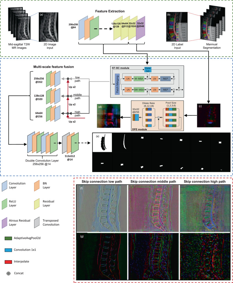
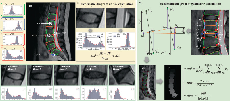
To help doctors and patients evaluate lumbar intervertebral disc degeneration (IVDD) accurately and efficiently, we propose a segmentation network and a quantitation method for IVDD from T2MRI. A semantic segmentation network (BianqueNet) composed of three innovative modules achieves high-precision segmentation of IVDD-related regions. A quantitative method is used to calculate the signal intensity and geometric features of IVDD. Manual measurements have excellent agreement with automatic calculations, but the latter have better repeatability and efficiency. We investigate the relationship between IVDD parameters and demographic information (age, gender, position and IVDD grade) in a large population. Considering these parameters present strong correlation with IVDD grade, we establish a quantitative criterion for IVDD. This fully automated quantitation system for IVDD may provide more precise information for clinical practice, clinical trials, and mechanism investigation. It also would increase the number of patients that can be monitored.


Conflict of interest statement

The authors declare no competing interests.

Figures

Fig. 1
Fig. 1. A flowchart of the study process from training and testing phase to data analysis phase with BianqueNet.
Mid-sagittal T2W lumbar MR images exported into two different resolutions (512*512, 320*320) are used to train and test two models (Model A, Model B). After segmentation accuracy evaluation, IVD parameters are quantified based on IVD-related area. As Model A shows good performance on quantitation consistency for MR images with various resolutions, it is used to establish baseline characteristic with different IVD degeneration among 5255 IVDs. Workflow diagram at the bottom presents the segmentation network, IVD quantitation method, and IVD degeneration determination.
Fig. 2
Fig. 2. The segmentation performance of BianqueNet in three typical cases and the influence of different segmentation accuracy on feature-point detection and calculation.
Segmentation results in case 1 and case 2 indicate that detailed information on the boundaries of vertebral bodies and IVDs is hard to detect. Segmentation in case 3 shows that irregular boundaries between the IVD and the vertebral bodies may interrupt segmentation with slight structural lesions or imaging defects. Feature-point-extracted results indicate that the precise segmentation may significantly improve the corner detection on vertebral bodies (red dots), thereby affecting the calculation results of the characteristic points on IVDs (green dots).
Fig. 3
Fig. 3. Baseline characteristics of IVD parameters in geometric and signal intensity.
The mean and standard deviation (σ) of the ΔSI of each of the modified Pfirrmann grading system (levels 1, 2, 3, 4, and 5) were calculated from Dataset C, which is used to quantify IVD degeneration (a), ΔSI (b), DH (c), DHI (d), and HDR (e) were quantified in different age, gender, and segments to establish population baseline.
Fig. 4
Fig. 4. The proposed BianqueNet consisted of three innovative modules.
a Input MRI, (b) annotations, feature map before (c) and after (d) DFE module, and (e) each image-channel output by the model corresponds to a segmentation area. Feature map of different skip-connection path with (f) and without (g) ST-SC module.
Fig. 5
Fig. 5. Scheme diagram of IVD parameter calculation.
Signal-intensity histogram calculation: (a) vertebral body area, (b) presacral fat area, (c) cerebrospinal fluid area, and (d) intervertebral disc area. e The outline of the segmented area is displayed on the original image. f Schematic diagram of ΔSI calculation. g Signal-intensity histogram corresponding to different Pfirrmann grade. h A geometric calculation method of lumbar disc-height parameters based on area, (i) vertebral body corner detection result (red points) and feature-point calculation result (green points), and (j) 80% area-extraction result of the intervertebral disc center.

References

    1. James SL, et al. Global, regional, and national incidence, prevalence, and years lived with disability for 354 Diseases and Injuries for 195 countries and territories, 1990-2017: A systematic analysis for the Global Burden of Disease Study 2017. Lancet. 2018;392:1789–1858. doi: 10.1016/S0140-6736(18)32279-7. - DOI - PMC - PubMed
    1. Urban JPG, Roberts S. Degeneration of the intervertebral disc. Arthritis Res. Ther. 2003;5:120–130. doi: 10.1186/ar629. - DOI - PMC - PubMed
    1. Hassan CR, Lee W, Komatsu DE, Qin YX. Evaluation of nucleus pulposus fluid velocity and pressure alteration induced by cartilage endplate sclerosis using a poro-elastic finite element analysis. Biomech. Model. Mechanobiol. 2021;20:281–291. doi: 10.1007/s10237-020-01383-8. - DOI - PMC - PubMed
    1. Khan AN, et al. Inflammatory biomarkers of low back pain and disc degeneration: a review. Ann. N. Y. Acad. Sci. 2017;1410:68–84. doi: 10.1111/nyas.13551. - DOI - PMC - PubMed
    1. Myers, E. R. & Wilson, S. E. Biomechanics of osteoporosis and vertebral fracture. Spine. 22, 25S–31S (1997). - PubMed

Publication types

MeSH terms