. 2022 Feb 11;12(1):2333.
doi: 10.1038/s41598-022-05207-w.

PLA/Hydroxyapatite scaffolds exhibit in vitro immunological inertness and promote robust osteogenic differentiation of human mesenchymal stem cells without osteogenic stimuli

Affiliations

PLA/Hydroxyapatite scaffolds exhibit in vitro immunological inertness and promote robust osteogenic differentiation of human mesenchymal stem cells without osteogenic stimuli

Marcela P Bernardo et al. Sci Rep. .

Abstract

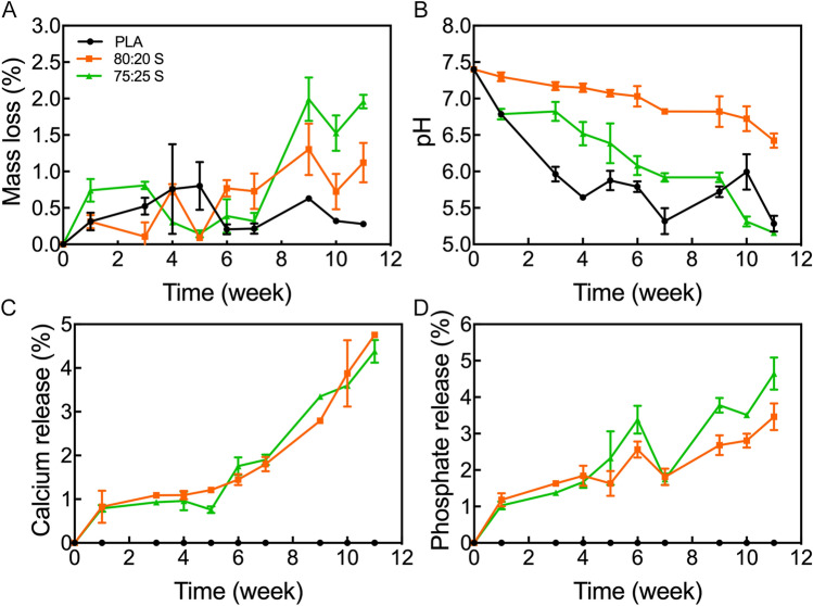
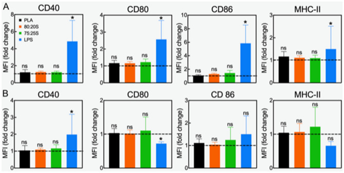
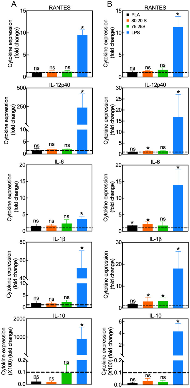
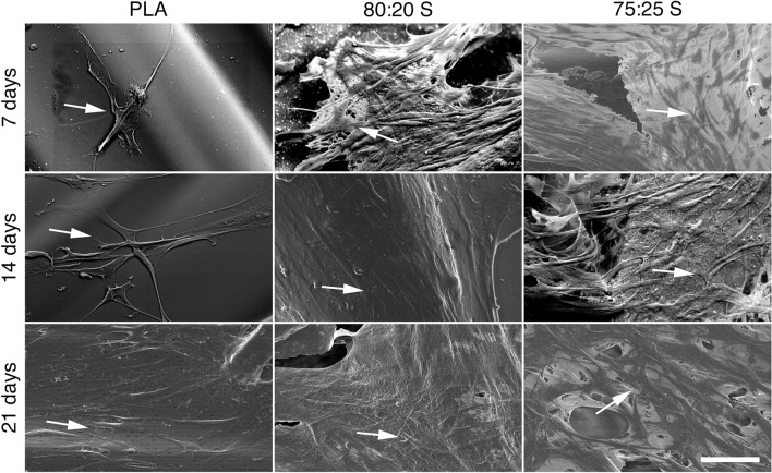
Bone defects stand out as one of the greatest challenges of reconstructive surgery. Fused deposition modelling (FDM) allows for the printing of 3D scaffolds tailored to the morphology and size of bone damage in a patient-specific and high-precision manner. However, FDM still suffers from the lack of materials capable of efficiently supporting osteogenesis. In this study, we developed 3D-printed porous scaffolds composed of polylactic acid/hydroxyapatite (PLA/HA) composites with high ceramic contents (above 20%, w/w) by FDM. The mechanical properties of the PLA/HA scaffolds were compatible with those of trabecular bone. In vitro degradation tests revealed that HA can neutralize the acidification effect caused by PLA degradation, while simultaneously releasing calcium and phosphate ions. Importantly, 3D-printed PLA/HA did not induce the upregulation of activation markers nor the expression of inflammatory cytokines in dendritic cells thus exhibiting no immune-stimulatory properties in vitro. Evaluations using human mesenchymal stem cells (MSC) showed that pure PLA scaffolds exerted an osteoconductive effect, whereas PLA/HA scaffolds efficiently induced osteogenic differentiation of MSC even in the absence of any classical osteogenic stimuli. Our findings indicate that 3D-printed PLA scaffolds loaded with high concentrations of HA are most suitable for future applications in bone tissue engineering.


Conflict of interest statement

The authors declare no competing interests.

Figures

Figure 1
Figure 1
In vitro analysis of (A) mass loss, (B) pH change, (C) calcium and (D) phosphate release for 3D-printed scaffolds. Error bars indicate one standard deviation above and below the means.
Figure 2
Figure 2
Surface markers expression of DC exposed to 3D-printed scaffolds for 24 (A) or 48 h (B). Results of two independent experiments using three-well replicates for each repetition are shown. Lines set to 1 represent the control (immature not activated DC). Ns no statistical difference; *p < 0.05. Please note that the statistical analysis refers to the comparison with the control.
Figure 3
Figure 3
Quantification of cytokine expression in DC incubated with 3D scaffolds for 24 (A) or 48 h (B). Data show the mean ± SD of two independent RT-PCR measurements. All values were normalized to the expression levels of GAPDH. Gene expression in control DC (cultured on tissue culture dishes) was set to one (dashed lines). LPS-treated DC served as the positive controls. *p < 0.05, ns non-significant. Please note that the statistical analysis refers to the comparison with the control.
Figure 4
Figure 4
SEM images showing the MSC adhesion on 3D-printed scaffolds after 7, 14 and 21 days of incubation. The arrows point to adherent MSC. Scale bar (for all panels): 50 μm.
Figure 5
Figure 5
Representative confocal microscopy images of MSC growing on PLA 3D-printed scaffold surface after 7 days of cultivation for (A) X–Y plane; (B) Y–Z plane; (C) X–Z plane. Cells were visualized with Alexa 647-conjugated phalloidin. The dashed line represents the surface of the scaffold. Direct imaging of PLA/HA 3D-printed scaffold was not possible due to their high intrinsic fluorescence.
Figure 6
Figure 6
(A) Evaluation of osteogenic differentiation of MSC on 3D-printed PLA/HA scaffolds using Alizarin Red staining. MSC were cultured on scaffolds for 21 days under growth conditions. Note the more intense Alizarin Red staining in the lower panels. (B) Quantification of Alizarin Red staining after elution and absorbance measurement at 405 nm. The line (set at 1) represents Alizarin Red staining level of control without scaffold. Error bars show one standard deviation above the mean. Scale bar (for all panels): 500 μm.
Figure 7
Figure 7
Flow cytometry analysis of CD10, CD105 and CD90 expression in MSC cultured on 3D-printed scaffold for 14 or 21 days under growth conditions. Dashed lines indicate surface marker expression (set to 1) in control cells cultured on tissue culture dishes. Error bars show one standard deviation above the mean.
Figure 8
Figure 8
Quantification of gene expression in MSC cultured on 3D scaffolds under growth conditions for 7, 14 or 21 days (AC, respectively). Data show the mean ± SD of two independent RT-PCR measurements. All values were normalized to GAPDH expression. Gene expression in control cells cultured on tissue culture dishes without scaffolds was set to one (dashed lines). *p < 0.05, ns non-significant. Please note that the statistical analysis refers to the comparison with the control.

References

    1. Turnbull G, et al. 3D bioactive composite scaffolds for bone tissue engineering. Bioact. Mater. 2018;3:278–314. - PMC - PubMed
    1. Corcione CE, Gervaso F, Madaghiele M, Sannino A, Licciulli A. Highly loaded hydroxyapatite microsphere/ PLA porous scaffolds obtained by fused deposition modelling. Ceram. Int. 2019;45:2803–2810.
    1. Nazeer, M. A. et al. 3D printed poly(lactic acid) scaffolds modified with chitosan and hydroxyapatite for bone repair applications. Mater. Today Commun.25, 101515 (2020).
    1. Farokhi M, et al. Importance of dual delivery systems for bone tissue engineering. J. Control. Release. 2016;225:152–169. - PubMed
    1. Hassanajili, S., Karami-Pour, A., Oryan, A. & Talaei-Khozani, T. Preparation and characterization of PLA/PCL/HA composite scaffolds using indirect 3D printing for bone tissue engineering. Mater. Sci. Eng. C104, 109960 (2019). - PubMed

Publication types