. 2022 Feb 11;12(1):2364.
doi: 10.1038/s41598-022-06228-1.

RYBP regulates Pax6 during in vitro neural differentiation of mouse embryonic stem cells

Affiliations

RYBP regulates Pax6 during in vitro neural differentiation of mouse embryonic stem cells

Enikő Sutus et al. Sci Rep. .

Abstract

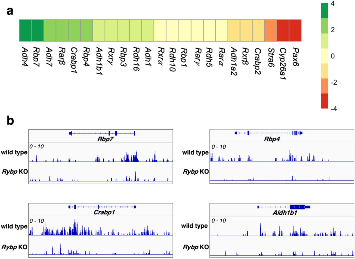
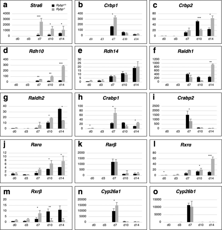
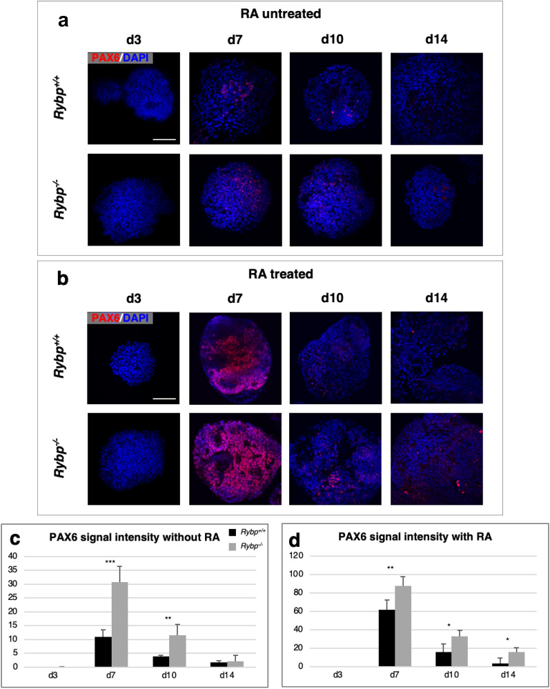
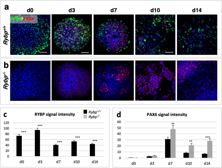
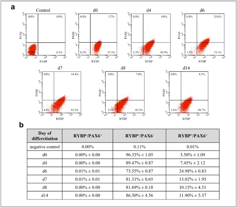
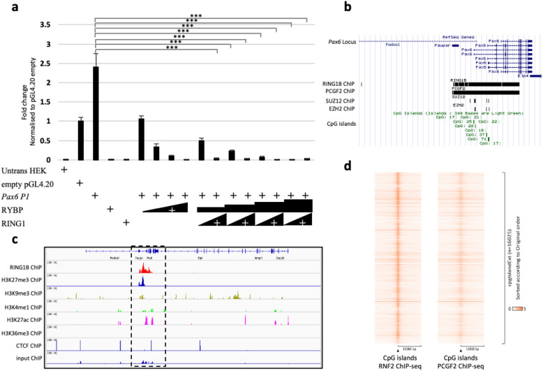
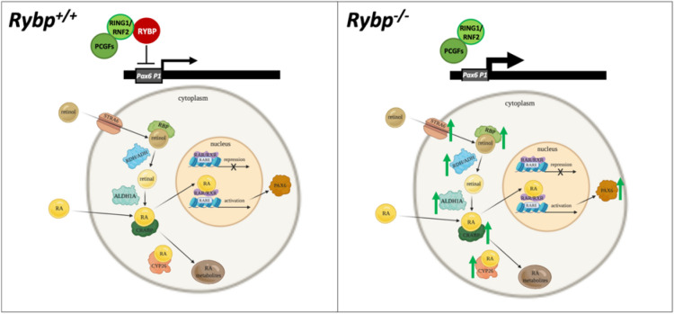
We have previously reported that RING1 and YY1 binding protein (RYBP) is important for central nervous system development in mice and that Rybp null mutant (Rybp-/-) mouse embryonic stem (ES) cells form more progenitors and less terminally differentiated neural cells than the wild type cells in vitro. Accelerated progenitor formation coincided with a high level of Pax6 expression in the Rybp-/- neural cultures. Since Pax6 is a retinoic acid (RA) inducible gene, we have analyzed whether altered RA signaling contributes to the accelerated progenitor formation and impaired differentiation ability of the Rybp-/- cells. Results suggested that elevated Pax6 expression was driven by the increased activity of the RA signaling pathway in the Rybp-/- neural cultures. RYBP was able to repress Pax6 through its P1 promoter. The repression was further attenuated when RING1, a core member of ncPRC1s was also present. According to this, RYBP and PAX6 were rarely localized in the same wild type cells during in vitro neural differentiation. These results suggest polycomb dependent regulation of Pax6 by RYBP during in vitro neural differentiation. Our results thus provide novel insights on the dynamic regulation of Pax6 and RA signaling by RYBP during mouse neural development.


Conflict of interest statement

The authors declare no competing interests.

Figures

Figure 1
Figure 1
Gene expression changes of RA signaling pathway members in wild type and Rybp−/− ES cells. (a) Heatmap of RA signaling related gene expression changes comparing Rybp−/− ES cells to wild type cells revealed altered expression of several RA pathway member genes. Green and red indicate upregulation (log2 fold change ≥ 1.5) and downregulation (log2 fold change ≤  − 1.5) respectively and yellow indicates genes that did not display significant expression changes. Heatmaps were generated using pheatmap (R package version 1.0.12) in Rstudio 1.3.959 ( http://www.rstudio.com/) (b) ChIP-seq analysis of H2AK119ub1 marks in wild type and Rybp−/− ES cells revealed loss of H2AK119ub1 at the genomic locus of RA pathway members. Data range 1–10.
Figure 2
Figure 2
Members of the RA signaling pathway exhibit elevated expression level in the absence of Rybp. Relative gene expression changes of membrane receptor (a) Stra6; intracellular retinol binding complex members (b) Rbp1, (c) Rbp2; and retinoid converting enzymes (d) Rdh10, (f) Aldh1a1, (g) Aldh1a2 by qRT-PCR showed altered expression in Rybp−/− neural cultures in comparison to wild type, while there was no significant difference in (e) Rdh14 expression in the two cell lines. Relative gene expression analysis of intracellular RA binding complex members (h) Crabp1, (i) Crabp2; nuclear RA receptor complex members (j) Rarα, (l) Rxrα, (m) Rxrβ and RA degrading enzyme (n) Cyp26a1 by qRT-PCR showed altered expression in the Rybp−/− neural cultures in comparison to the wild type but there was no difference in (k) Rarβ and (o) Cyp26b1 expression between the two cell lines. The expression of the indicated markers was normalized to Hprt level. Means are standard deviation ± SD. Values of p < 0.05 were accepted as significant (*p < 0.05; **p < 0.01; ***p < 0.001). Statistical method: t test type 3, n = 3.
Figure 3
Figure 3
PAX6 is strongly upregulated in the lack of Rybp (Rybp−/−) during neural differentiation of ES cells. Immunocytochemical analysis of PAX6 protein level in EBs in the (a) absence and (b) presence of atRA revealed increased PAX6 signals in the Rybp−/− cells compared to the wild type cells. Objective: 40 × . Scale bar: 80 μm. (c,d) ImageJ quantification of PAX6 protein level in the wild type and Rybp−/− cells during the in vitro neural differentiation in the absence (c) and presence (d) of atRA. Means are standard deviation ± SD. Values of p < 0.05 were accepted as significant (*p < 0.05; **p < 0.01; ***p < 0.001). Statistical method: t test type 3, n = 3.
Figure 4
Figure 4
Immunocytochemical localization of RYBP and PAX6. Immunocytochemical analysis of RYBP and PAX6 protein level in (a) Rybp+/+ and (b) Rybp−/− cells during in vitro neural differentiation. Objective: d0, d10 and d14: 60x; d3 and d7: 40x. Scale bar: d0, d10 and d14: 100 μm; d3 and d7: 80 μm. (c,d) ImageJ quantification of (c) RYBP and (d) PAX6 protein level in the wild type and Rybp−/− cells during in vitro neural differentiation. Means are standard deviation ± SD. Values of p < 0.05 were accepted as significant (*p < 0.05; **p < 0.01; ***p < 0.001). Statistical method: t test type 3, n = 3.
Figure 5
Figure 5
RYBP and PAX6 are co-expressed in cells during in vitro neural differentiation. The wild type ES cells were induced for differentiation with atRA for various periods of time (d4, d6, d7, d8 and d14) or left unstimulated (d0). The cells were stained with mouse monoclonal PAX6 and rabbit polyclonal RYBP/DEDAF primary antibodies followed with Alexa Fluor 647 labelled Donkey anti Mouse IgG (H + L) and Alexa Fluor 488 labelled Donkey anti Rabbit IgG (H + L), respectively then analyzed with cytofluorimetry. Control means, that the first antibodies were omitted and both secondary antibodies were added to the samples (a,b). Representative FACS plots of two independent experiments is shown at (a). Data are represented as means me of two biological repeats (n = 2). (b) Percentage and standard deviation ( ±) of control, RYBP/PAX6+, RYBP+/PAX6 and RYBP+/PAX6+ cells during in vitro neural differentiation of the wild type ES cells. Percentages were calculated with BD CellQuest Pro Version 6.0 software.
Figure 6
Figure 6
RYBP represses Pax6 gene expression through the Pax6 P1 promoter. (a) The Pax6 genomic locus showing the position of the 2014 bp P1 promoter used in this study. (b) CpG island prediction at the Pax6 P1 promoter was performed in DBCAT (http://dbcat.cgm.ntu.edu.tw). The predicted CpG island is represented in blue and the denser CpG regions are displayed in yellow. (c) Luciferase assays were performed in HEK293 cells transiently transfected with mouse Pax6 P1 promoter reporter (pGL4.20 Pax6 P1) or with empty vector (pGL4.20) and RYBP expression plasmid in increasing dosages (1 µg, 5 µg, 10 µg and 20 µg). Values are expressed as fold changes of luciferase activity normalized to the empty vector. The presented values are averages of three independent experiments. Means are standard deviation ± SD. Values of p < 0.05 were accepted as significant (*p < 0.05; **p < 0.01; ***p < 0.001, n = 3). Statistical method: T test type 3. (d) Pax6 P1 promoter highlighting the position of key transcription factor binding sites and CpG islands. CpG island is represented in italics. The transcription factor binding sites of RYBP interacting proteins are highlighted in grey. PAX6 self-binding sites are represented in bold, RXRα and SP1 binding sites are underlined. Untrans HEK Untransfected HEK293 cells, SP1 Trans-acting transcription factor 1, E2F E2 transcription factor, MYC/MAX Myelocytomatosis oncogene/MYC associated factor X, YY1 Yin-yang-1 transcription factor.
Figure 7
Figure 7
RYBP represses Pax6 gene expression in a polycomb dependent way. (a) Luciferase assays were performed in HEK293 cells transiently transfected with mouse Pax6 P1 promoter reporter (pGL4.20 Pax6 P1) or with empty vector (pGL4.20), RYBP and RING1 expression plasmids in increasing dosages (Rybp: 1 µg, 5 µg, 10 µg, 20 µg; Ring1: 1 µg, 10 µg) Values are expressed as fold changes of luciferase activity normalized to the empty vector. The presented values are averages of three independent experiments. Means are standard deviation ± SD. Values of p < 0.05 were accepted as significant (*p < 0.05; **p < 0.01; ***p < 0.001, n = 3). Statistical method: t test type 3. (b) UCSC genome tracks showing ChIP-seq binding of PRC1 factors RING1 (GSM1917303), PCGF2 (GSM1917304), and PRC2 factors SUZ12 (GSM1917301) and EZH2 (GSM1917302). (c) The methylation state of Pax6 genomic locus in NPCs determined by the histone modifications present using existing ChIP-seq data from Bonev et al., 2017. The binding of RNF2 (GSM2533860), H3K27me3 (GSM2533864), H3K9me3 (GSM2533866), H3K4me1 (GSM2533862), H3K27ac (GSM2533868) and H3K36me3 (GSM2533870) are displayed in reference to CTCF (GSM2533858) and input (GSM2533871). The Pax6 P1 promoter (highlighted in dotted box) displayed bivalency presenting both repression histone marks (H3K27me3 and H3K9me3) and activation histone mark (H3K27ac) in NPCs. (d) The peak density heat map of the genome wide binding targets of RNF2 and PCGF2 integrated with the global position of the CpG islands in NPCs. Untrans HEK Untransfected HEK293 cells.
Figure 8
Figure 8
RYBP directs neurogenesis through the regulation of RA pathway and Pax6. In the absence of Rybp the mRNA expression of the RA pathway members and RYBP target gene Pax6 are upregulated. Normal Rybp level is required to the normal neural differentiation. RYBP can regulate the Pax6 expression via ncPRC1. The green arrows represent the upregulation of the gene expression of the RA pathway members.

Similar articles

Cited by

References

    1. Chuang J, Tung L, Lin Y. Neural differentiation from embryonic stem cells in vitro: An overview of the signaling pathways. World J. Stem Cells. 2015;7:437. - PMC - PubMed
    1. Tonge PD, Andrews PW. Retinoic acid directs neuronal differentiation of human pluripotent stem cell lines in a non-cell-autonomous manner. Differ. Res. Biol. Divers. 2010;80:20–30. - PubMed
    1. Bonney S, et al. Diverse functions of retinoic acid in brain vascular development. J. Neurosci. 2016;36:7786–7801. - PMC - PubMed
    1. Gajović S, St-Onge L, Yokota Y, Gruss P. Retinoic acid mediates Pax6 expression during in vitro differentiation of embryonic stem cells. Differ. Res. Biol. Divers. 1997;62:187–192. - PubMed
    1. Rhinn M, Dollé P. Retinoic acid signalling during development. Development. 2012;139:843–858. - PubMed

Publication types

MeSH terms