Review
. 2022 Apr;22(4):239-252.
doi: 10.1038/s41568-022-00444-7. Epub 2022 Feb 11.

Liquid-liquid phase separation drives cellular function and dysfunction in cancer

Affiliations
Review

Liquid-liquid phase separation drives cellular function and dysfunction in cancer

Sohum Mehta et al. Nat Rev Cancer. 2022 Apr.

Abstract

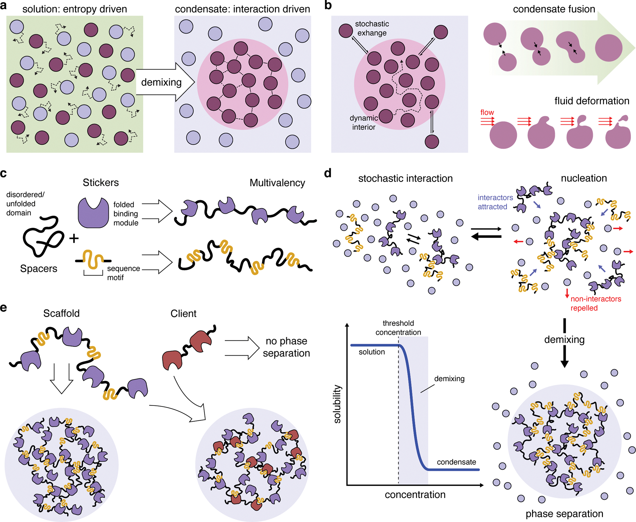
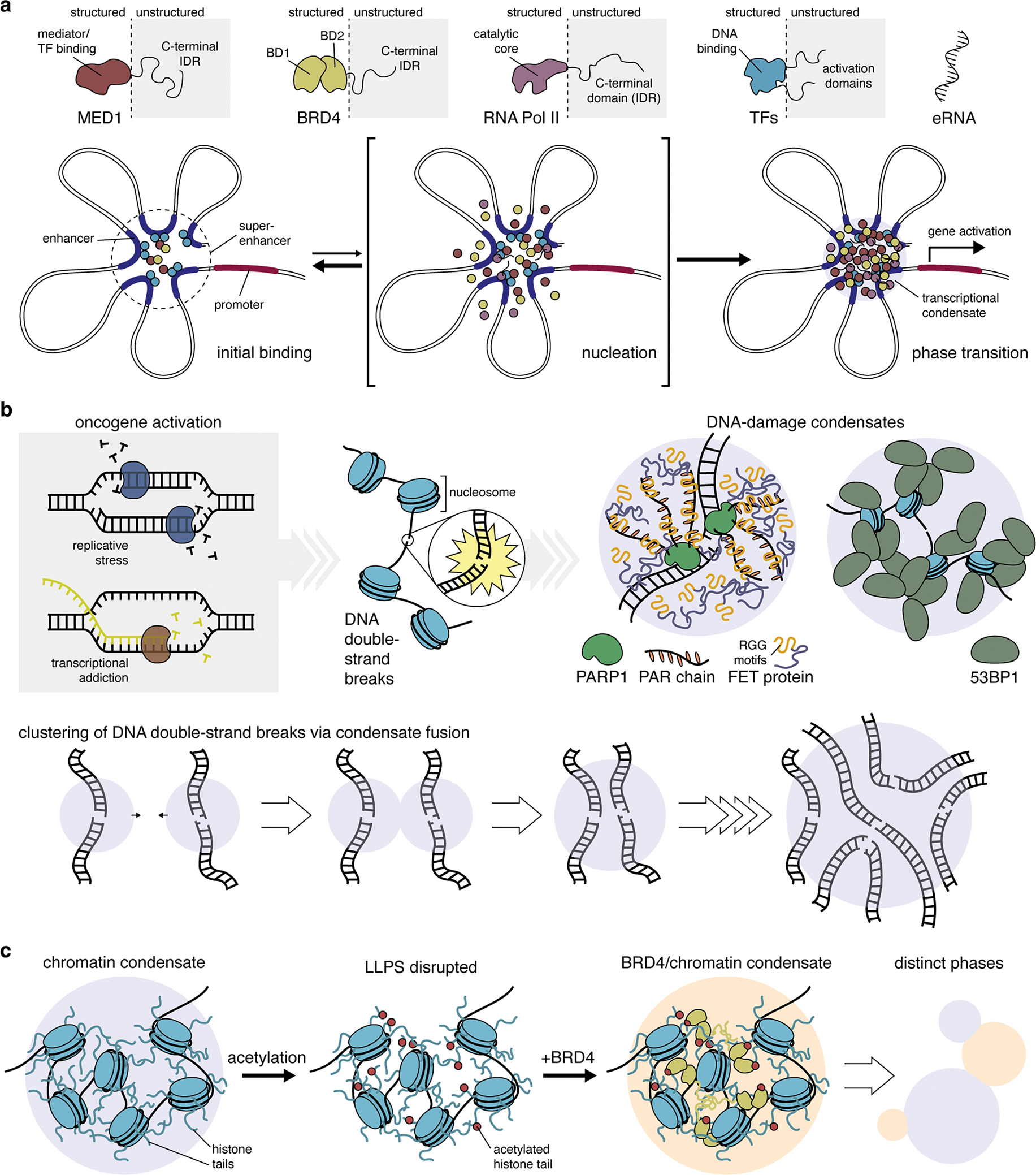
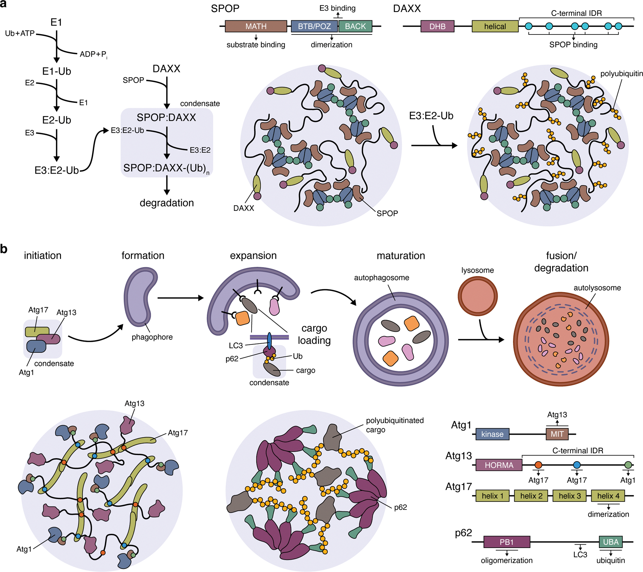
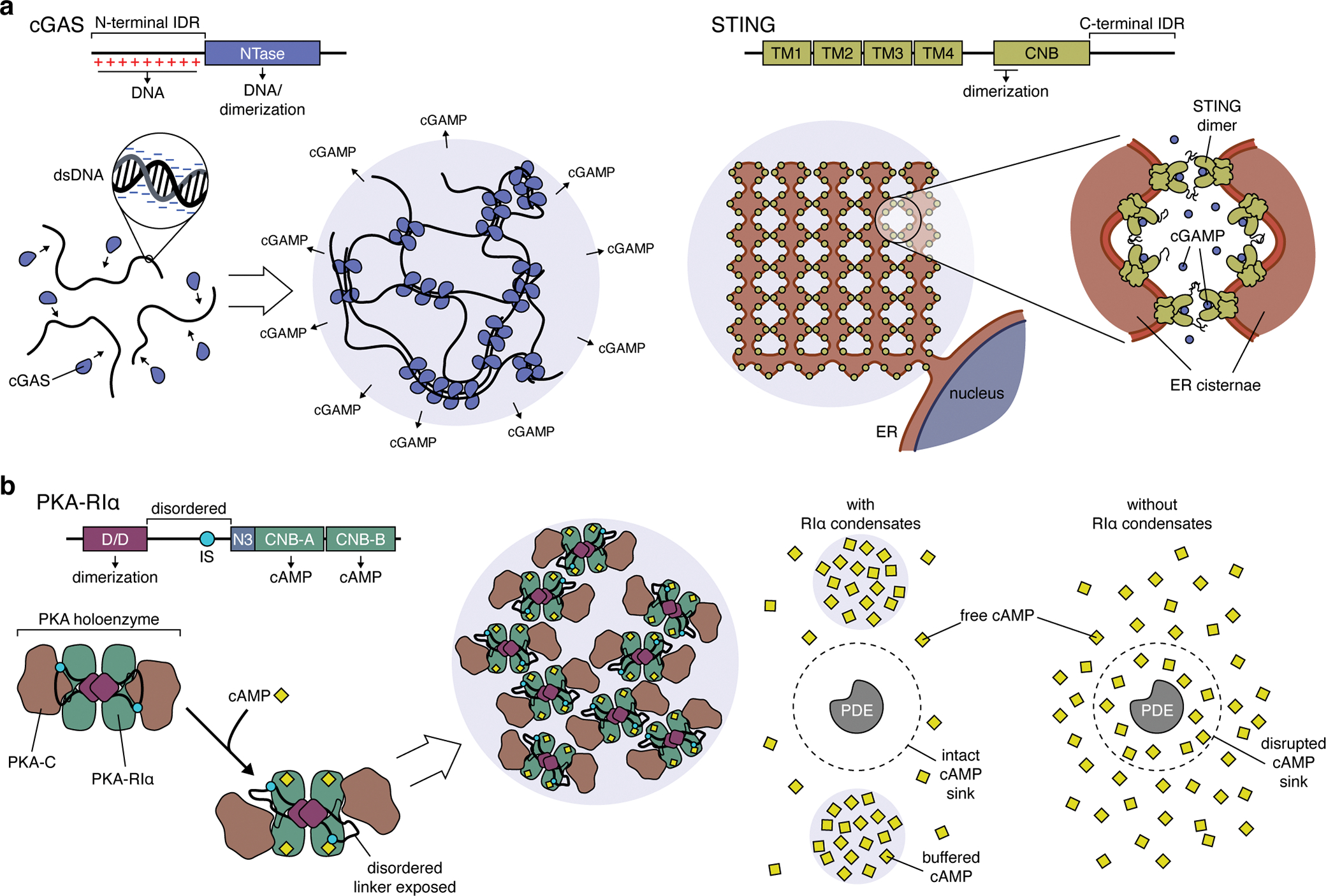
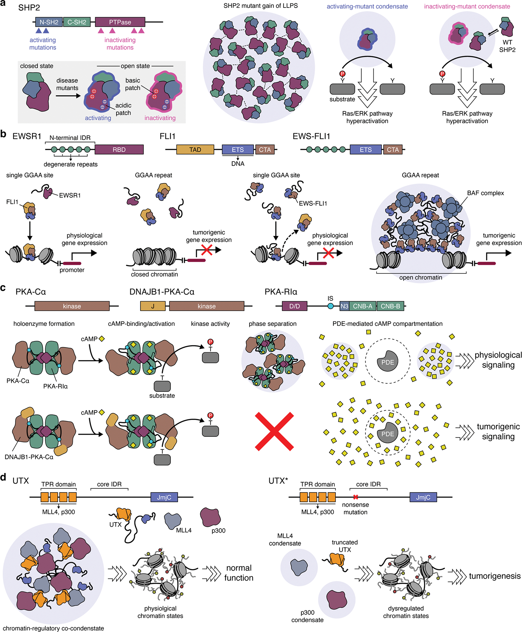
Cancer is a disease of uncontrollably reproducing cells. It is governed by biochemical pathways that have escaped the regulatory bounds of normal homeostatic balance. This balance is maintained through precise spatiotemporal regulation of these pathways. The formation of biomolecular condensates via liquid-liquid phase separation (LLPS) has recently emerged as a widespread mechanism underlying the spatiotemporal coordination of biological activities in cells. Biomolecular condensates are widely observed to directly regulate key cellular processes involved in cancer cell pathology, and the dysregulation of LLPS is increasingly implicated as a previously hidden driver of oncogenic activity. In this Perspective, we discuss how LLPS shapes the biochemical landscape of cancer cells.


Conflict of interest statement

COMPETING INTERESTS

The authors declare they have no competing interests.

Figures

Figure 1.
Figure 1.. Basic principles of liquid-liquid phase separation and biomolecular condensates.
(a) Whereas entropy typically drives molecules to become dispersed in solution, mutual interactions among a subset of molecules can shift the free-energy landscape to favor de-mixing and drive the formation of a separate condensed phase. (b) Biomolecular condensates display liquid-like properties, such as highly dynamic interiors capable of rapid rearrangement and stochastic exchange with the bulk solution, as well as dynamic fusion, deformation, and fission events. (c) Phase separation of biomolecules is driven my multivalent binding interactions. These molecules often exhibit a sticker-and-spacer configuration, in which multiple folded binding modules and/or short interaction sequence (stickers) are separated by unfolded or disordered regions (spacers), thus achieving multivalency while promoting liquid-like behaviors. (d) At low concentrations, molecules capable of phase-separation interact randomly but remain in solution. Increasing the concentration of these molecules promotes nucleation of small complexes, which begin to exclude the surrounding solution. Each new interactor contributes binding sites that attract more interactors. This positive feedback loop results in switch-like de-mixing and phase separation (blue curve) once a critical threshold concentration is reached. (e) Phase-separating biomolecules are often categorized as scaffolds, which are both necessary and sufficient for phase separation, and clients, which can selectively partition into scaffold-containing condensates but cannot independently phase separate.
Figure 2.
Figure 2.. The regulation of nuclear function by liquid-liquid phase separation.
(a) Many transcriptional regulators, such as mediator complex subunit 1 (MED1), bromodomain-containing protein 4 (BRD4), RNA polymerase II (RNA Pol II), and various transcription factors (TFs) feature large intrinsically disordered regions (IDRs) that allow them to undergo phase separation and form biomolecular condensates at super-enhancer sites. Initial binding of TFs and other regulatory proteins at neighboring enhancer sites triggers the nucleation of additional factors via multivalent interactions mediated by their IDRs. This short-lived intermediate rapidly gives rise to a phase transition, yielding a transcriptional condensate that drives gene activation. Thus, LLPS is thought to play a key role in the robust, switch-like regulation of cell-identity genes by super-enhancers. (b) High levels of replicative stress and excessive transcription induced by oncogene activation cause the accumulation of genome damage, including DNA double-strand breaks (DSBs). The binding of poly-(ADP-ribose) polymerase 1 (PARP1) and synthesis of PAR chains at DSB sites leads to the recruitment of disordered FET-family proteins via their RGG repeats, resulting in the formation of phase-separated condensates. Similarly, P53 binding protein 1 (53BP1) binds nucleosome proteins near DSB sites, resulting in the formation of phase-separated condensates via 53BP1 oligomerization. Notably, 53BP1 condensates have been observed to fuse, suggesting a possible mechanism of the clustering of DSBs in cells (lower panel). (c) Chromatin undergoes LLPS as a result of multivalent interactions driven by intrinsically disordered histone tails. LLPS of chromatin can be modulated by post-translational modification of histone tails, such as acetylation, which disrupts condensate formation. The addition of BRD4, which binds acetylated histone tails, restores LLPS of acetylated chromatin. The resulting condensates cannot mix with unmodified chromatin condensates, yielding two distinct phases, similar to distinct chromatin domains in the nucleus.
Figure 3.
Figure 3.. The role of liquid-liquid phase separation in cellular quality control.
(a) The formation of biomolecular condensates between speckle-type POZ protein (SPOP) and its target death-domain-associated protein (DAXX). Left: Pathway schematic highlighting the role of SPOP:DAXX condensates in facilitating DAXX polyubiquitination. Right: LLPS of SPOP and DAXX is driven by the formation of SPOP dimers through its BR-C/ttk/bab (BTB)/Pox virus and zinc finger (POZ) domain, interactions between SPOP dimers mediated by their BTB and C-terminal Kelch (BACK) domains, as well as interactions between the long, C-terminal IDR of DAXX with multiple copies of the N-terminal meprin and TRAF homology (MATH) domain of SPOP. The BTB/POZ-BACK domain junction in SPOP also binds E3 ubiquitin ligases, recruiting them into condensates (omitted from cartoon) to promote DAXX polyubiquitination. Ub, ubiquitin; (Ub)n, polyubiquitin; E1, Ub-activating enzyme; E2, Ub-conjugating enzyme; E3, Ub-ligase. (b) Top: Simplified schematic of the autophagy pathway, highlighting the involvement of Atg1, Atg13, and Atg17 condensates in autophagy initiation and the role of p62 condensates in cargo loading during phagophore expansion. Bottom: Atg1:Atg13:Atg17 condensates (left panel) are scaffolded by Atg13 and Atg17, with the C-terminal IDR of Atg13 containing a pair of binding sites that crosslink Atg17 dimers. Atg1 is recruited as a client protein via its C-terminal microtubule-interacting and transport (MIT) domain, which binds Atg13. Bottom: LLPS of p62 condensates (middle panel) requires the formation of p62 oligomers, mediated by an N-terminal Phox and Bem1p (PB1) domain, whose C-terminal ubiquitin-associated (UBA) domains then participate in multivalent interactions with the ubiquitin chains on polyubiquitinated substrate proteins.
Figure 4.
Figure 4.. Liquid-liquid phase separation shapes signaling pathway function.
(a) Left: cGAS binds double-stranded DNA (dsDNA) via its positively charged N-terminal IDR and its C-terminal nucleotidyltransferase (NTase) domain, which also mediates cGAS dimerization. These multivalent interactions rapidly nucleate the formation of cGAS:dsDNA condensates, dramatically lowering the concentration threshold for cGAS activation and leading to ultrasensitive dsDNA detection and cGAMP production. Right: However, excess cGAMP production has been shown to trigger LLPS of the ER-resident cGAMP receptor STING, which requires interactions mediated by the C-terminal IDRs in STING dimers and forms condensates containing intricately folded membrane networks. Because LLPS requires cGAMP-bound STING, these condensates may function to buffer excess cGAMP and regulate immune signaling. (b) Left: The formation of PKA-RIα biomolecular condensates requires the docking and dimerization (D/D) domain and a disordered linker region containing an inhibitory site (IS) that binds the PKA-C subunit. The binding of cAMP to RIα triggers a conformational rearrangement in the PKA holoenzyme that exposes the disordered linker to help drive LLPS, though the precise molecular mechanism remains unclear. Right: RIα condensates dynamically buffer cAMP levels, allowing PDEs to efficiently degrade free cAMP and act as cAMP sinks. In the absence of RIα condensates, PDEs catalytic activity is overwhelmed, resulting in the collapse of cAMP sinks and the breakdown of compartmentalized cAMP signaling.
Figure 5.
Figure 5.. Dysregulation of liquid-liquid phase separation as a tumorigenic driver.
(a) Gain of LLPS ability by disease-causing SHP2 mutants. Both activating and inactivating mutations in SHP2 are able to trigger oncogenic signaling by altering SHP2 conformation to favor the open state. This conformation exposes a patch of positively charged, basic residues on the surface of the SHP2 protein tyrosine phosphatase (PTPase) domain, which can then form electrostatic interactions with negatively charged acidic residues elsewhere on the protein surface, thus driving LLPS by these mutants. Condensates formed by activating SHP2 mutants induce Ras/ERK pathway hyperactivation through their intrinsically high PTPase activity. By contrast, while condensates formed by inactivating SHP2 mutants gain PTPase activity by recruiting and activating wild-type (WT) SHP2 to trigger hyperactive signaling. (b) Gain of LLPS by EWS-FLI1 oncogenic fusion. In Ewing sarcoma, the N-terminal IDR of the EWSR1 becomes fused to the C-terminal DNA-binding domain of FLI1. Left: Native FLI1 binds conserved enhancer sites containing a single GGAA consensus sequence to control physiological gene expression but is unable to bind GGAA microsatellite repeats. Right: The EWS-FLI1 fusion disrupts the binding of native FLI1 to enhancers and also gains the ability to undergo LLPS via its IDR. EWS-FLI1 condensates tightly bind GGAA microsatellites while also strongly recruiting the BAF chromatin remodeling complex, producing open chromatin sites that act as super-enhancers for pro-tumor gene expression. (c) Pathological loss of LLPS by an oncogenic fusion. In the DNAJB1-PKA-Cα chimera, an N-terminal portion of DNAJB1 encompassing the J-domain (J) replaces the native N-terminus of WT PKA-Cα. WT PKA-Cα and DNAJB1-PKA-Cα behave similarly in forming holoenzymes with PKA-R subunits (e.g., RIα), in undergoing cAMP-induced activation, and in phosphorylating substrates. However, DNAJB1-PKA-Cα completely abolishes RIα LLPS, which disrupts the compartmentation of cAMP signaling by PDEs, leading to tumorigenic signaling. (d) Loss of LLPS by UTX mutants disrupts chromatin regulation. Left: In healthy cells, histone demethylase UTX undergoes LLPS mediated by a core IDR between the tetratricopeptide repeat (TPR) and Jumonji C (JmjC) domains. The TPR domain of UTX recruits the MLL4 histone methyl transferase and the p300 histone acetyl transferase to form chromatin-regulating co-condensates that maintain physiological chromatin states. Right: UTX is frequently mutated in cancers, with the most frequent alteration being a nonsense mutation near the start of the core IDR. These mutant UTX variants (UTX*) cannot phase separate with MLL4 and p300, leading to dysregulated chromatin states and tumorigenesis.

References

    1. Lengauer C, Kinzler KW & Vogelstein B Genetic instabilities in human cancers. Nature 396, 643–649 (1998). - PubMed
    1. Sever R & Brugge JS Signal Transduction in Cancer. Csh Perspect Med 5, a006098 (2015). - PMC - PubMed
    1. Sanchez-Vega F et al. Oncogenic Signaling Pathways in The Cancer Genome Atlas. Cell 173, 321–337.e10 (2018). - PMC - PubMed
    1. Bradner JE, Hnisz D & Young RA Transcriptional Addiction in Cancer. Cell 168, 629–643 (2017). - PMC - PubMed
    1. Sengupta S & George RE Super-Enhancer-Driven Transcriptional Dependencies in Cancer. Trends Cancer 3, 269–281 (2017). - PMC - PubMed

Publication types