. 2022 Jun;29(30):45751-45762.
doi: 10.1007/s11356-022-19006-3. Epub 2022 Feb 11.

Reproductive effects of sulfoxaflor in male Sprague Dawley rats

Affiliations

Reproductive effects of sulfoxaflor in male Sprague Dawley rats

Wafaa H Mohamed et al. Environ Sci Pollut Res Int. 2022 Jun.

Abstract

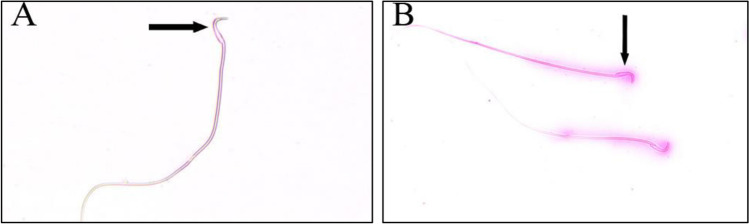
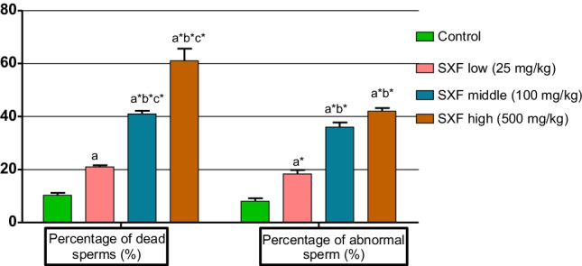
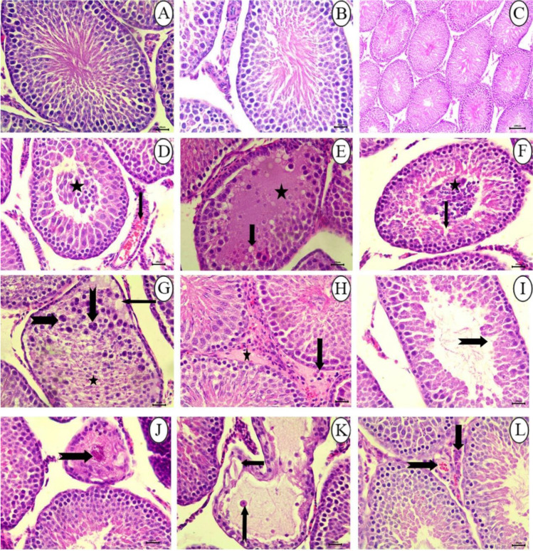
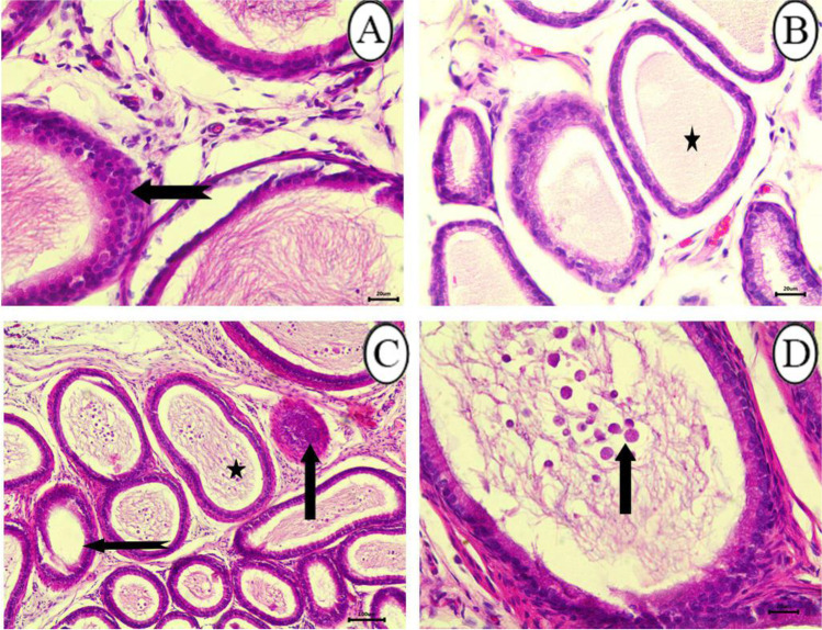
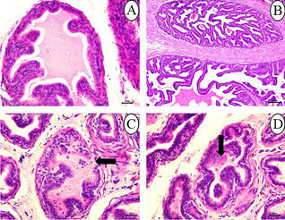
The study objective was to evaluate the potential reproductive toxicity of sulfoxaflor (SFX) insecticide in male Sprague Dawley rats. To attain these objectives, forty male Sprague Dawley rats of 10-12 weeks old were randomly divided into four equal groups; the 1st group was used as a control group; the other three groups were exposed to 25, 100, and 500 mg/kg body weight SFX by oral gavage for 4 weeks. Relative testicular weight, testosterone, FSH, LH, MDA, and GPx levels, sperm viability, sperm morphology, sperm DNA damage, and histopathological changes in testes, epididymis, and seminal vesical of these rats were investigated after 4 weeks. The results showed that SFX exposure resulted in a significant increase in FSH, LH, MDA, and GPx levels as well as the percentage of dead and abnormal sperms and DNA damage in rat sperms. Histopathological examination of testes established testicular degeneration with coagulative necrosis as well as the proliferation of interstitial connective tissue infiltrated with inflammatory cells with congestion of intertubular blood vessels in epididymis and degeneration of lining epithelium of seminal vesicles.

Keywords: Reproductive toxicity; Sperm morphology and sperm DNA damage; Sperm viability; Sulfoxaflor.


Conflict of interest statement

The authors declare no competing interests.

Figures

Fig. 1
Fig. 1
A Eosine nigrosin-stained semen film of control rats showed normal live epididymal sperm, not stained (arrow). B Semen film of SFX-treated rats showed dead epididymal sperms (appeared pink) (arrow) (× 40)
Fig. 2
Fig. 2
Photos of methyl violet stained semen film of control and SFX-treated rats showing epididymal sperms morphology. (A) Normal sperm, (B) detached head, (C) broken tail, (D) microcephalic head, (E) double head with a single tail, and (F) coiled tail (× 40)
Fig. 3
Fig. 3
The effect of SFX on sperm viability and morphology in male rats exposed to 25, 100, and 500 mg/kgb.w. of SFX for 4 weeks. * indicates highly significance at p ≤ 0.01; a indicates significance at p ≤ 0.05 in comparison with the control group; b Indicates significance at p ≤ 0.05 in comparison with 25 mg/kg dose group; c indicates significance at p ≤ 0.05 between 100 and 500 mg/kg dose groups
Fig. 4
Fig. 4
A Acridine orange-stained semen film of SFX-exposed rats showed damaged sperm DNA (yellow head). B Semen film of control rats showed normal intact sperm DNA (appeared green head) (× 40)
Fig. 5
Fig. 5
Histopathological examination of testes in (A and B) control group showing seminiferous tubules lined by normal germinal epithelium, bar = 20 μm; (C) control group showing seminiferous tubules, bar = 100 μm; (D) 25 mg/kg SFX-exposed group showing a collection of tissue debris inside the lumen of seminiferous tubules (star) and hyperemia (arrow), bar = 20 μm; (E) 25 mg /kg SFX-exposed group showing necrosis of germinal epithelium (arrow) and lumen filled with oedematous fluid (star), bar = 20 μm; (F) 100 mg/kg SFX-exposed group showing (spermatid giant cells) (star), degeneration of germinal epithelium (arrow), bar = 20 μm; (G) 100 mg/kg SFX-exposed group showing hypereosinophilic disorganized cells (star), vacuolation (arrow), and formation of (spermatid giant cells) (notched arrow), bar = 20; (H) 100 mg/kg SFX-exposed group showing interstitial oedema (star) with mild hyperplasia of (Leydig cells) (arrow), bar = 20 μm; (I) 100 mg/kg SFX-induced group showing necrosis of germinal epithelium (notched arrows), bar = 20 μm; (J) 500 mg/kg SFX-induced group showing coagulative necrosis in germinal epithelium (notched arrow), bar = 20 μm; (K) 500 mg/kg SFX-exposed group showing detachment of germinal epithelium (arrow) with the presence of eosinophilic fluid in the lumen of seminiferous tubules (star) and multinucleated giant cells (notched arrow), bar = 20; and (L) 500 mg/kg SFX-exposed group showing congestion of interstitial blood vessels (notched arrow) and hyperplasia of (Leydig cells) (arrow), bar = 20 μm, H&E
Fig. 6
Fig. 6
Histopathological examination of epididymis in (A) control group showing normal epididymal convoluted tubule lined with pseudostratified columnar epithelium bar = 20 μm; (B) control group showing epididymal ducts in, bar = 100 μm; (C) 25 mg/kg SFX-exposed group showing decrease in number of sperms inside the epididymal ducts (star), bar = 100 μm; and (D) 25 mg/kg SFX-exposed group showing hyperemia (arrow), proliferation of interstitial connective tissue (star), bar = 20 μm, H&E
Fig. 7
Fig. 7
Histopathological examination of epididymis in (A) 100 mg/kg SFX-exposed group showing hyperplasia of lining epithelium (notched arrow), bar = 20 μm; (B) 100 mg/kg SFX-exposed group showing that the most of epididymal ducts were devoid from spermatozoa (star), bar = 20 μm; (C) 500 mg/kg SFX-exposed group showing accumulation of (spermatid giant cells) (star), epididymal duct completely free from spermatozoa (notched arrow), and complete necrosis of epididymal duct (arrow), bar = 100 μm; and (D) 500 mg/kg SFX-exposed group showing accumulation of (spermatid giant cells) (notched arrow); bar = 20 μm, H&E
Fig. 8
Fig. 8
Histopathological examination of seminal vesicles in (A) control group showing folded mucosa lined with secretory epithelium, bar = 20 μm; (B) the seminal vesicles in control group, bar = 100 μm; (C) 25 mg/kg SFX-exposed group showing degeneration and sloughing of lining epithelium (arrow), bar = 20 μm; (D) 25 mg/kg SFX-exposed group showing hyperplasia of lining epithelium (arrows), bar = 20 μm, H&E
Fig. 9
Fig. 9
Histopathological examination of seminal vesicles in (A) 100 mg/kg SFX-exposed group showing papillary structure extending into the glandular lumen (arrow), bar = 20 μm; (B) 100 mg/kg SFX-induced group showing increased prominence of the contracted fibromuscular stroma (arrow), bar = 100 μm; (C) 500 mg/kg SFX-exposed group showing hyperplasia of lining epithelium (arrow), connective tissue proliferation in lamina propria (star), and hemorrhage (notched arrow), bar = 20 μm, H&E

References

    1. Airaodion AI, Ngwogu AC, Megwas AU, Ekenjoku JA, Ngwogu KO. Effect of common household insecticides used in Nigeria on rat male reproductive hormones. Int J Res Rep in Gynaecol. 2019;2(1):1–8.
    1. Ali RI, Ibrahim MA. Malathion induced testicular toxicity and oxidative damage in male mice: the protective effect of curcumin. Egypt J Forensic Sci. 2018;8:1–13. doi: 10.1186/s41935-018-0099-x. - DOI
    1. Arıcan EY, Kayalı DG, Karaca BU, Boran T, Öztürk N, Okyar A, Ercan F, Özhan G. Reproductive effects of subchronic exposure to acetamiprid in male rats. J Scientific Reports. 2020;10:1–10. doi: 10.1038/s41598-019-56847-4. - DOI - PMC - PubMed
    1. Authority E F S. Crivellente F, Hart A, Hernandez-Jerez AF, Bennekou SH, Pedersen R, Terron A, Wolterink G, Mohimont L. Establishment of cumulative assessment groups of pesticides for their effects on the nervous system. EFSA J. 2019;17(9):5800–5115. - PMC - PubMed
    1. Bacci L, Convertini S, Rossaro B. A review of sulfoxaflor, a derivative of biological acting substances as a class of insecticides with a broad range of action against many insect pests. J Entomol Acarol Res. 2018;50(3):7836. doi: 10.4081/jear.2018.7836. - DOI

LinkOut - more resources