. 2022 Feb 12;12(1):13.
doi: 10.1186/s13613-022-00988-9.

Sequential lateral positioning as a new lung recruitment maneuver: an exploratory study in early mechanically ventilated Covid-19 ARDS patients

Affiliations

Sequential lateral positioning as a new lung recruitment maneuver: an exploratory study in early mechanically ventilated Covid-19 ARDS patients

Rollin Roldán et al. Ann Intensive Care. .

Abstract

Background: A sequential change in body position from supine-to-both lateral positions under constant ventilatory settings could be used as a postural recruitment maneuver in case of acute respiratory distress syndrome (ARDS), provided that sufficient positive end-expiratory pressure (PEEP) prevents derecruitment. This study aims to evaluate the feasibility and physiological effects of a sequential postural recruitment maneuver in early mechanically ventilated COVID-19 ARDS patients.

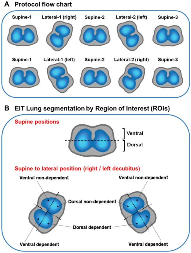
Methods: A cohort of 15 patients receiving lung-protective mechanical ventilation in volume-controlled with PEEP based on recruitability were prospectively enrolled and evaluated in five sequentially applied positions for 30 min each: Supine-baseline; Lateral-1st side; 2nd Supine; Lateral-2nd side; Supine-final. PEEP level was selected using the recruitment-to-inflation ratio (R/I ratio) based on which patients received PEEP 12 cmH2O for R/I ratio ≤ 0.5 or PEEP 15 cmH2O for R/I ratio > 0.5. At the end of each period, we measured respiratory mechanics, arterial blood gases, lung ultrasound aeration, end-expiratory lung impedance (EELI), and regional distribution of ventilation and perfusion using electric impedance tomography (EIT).

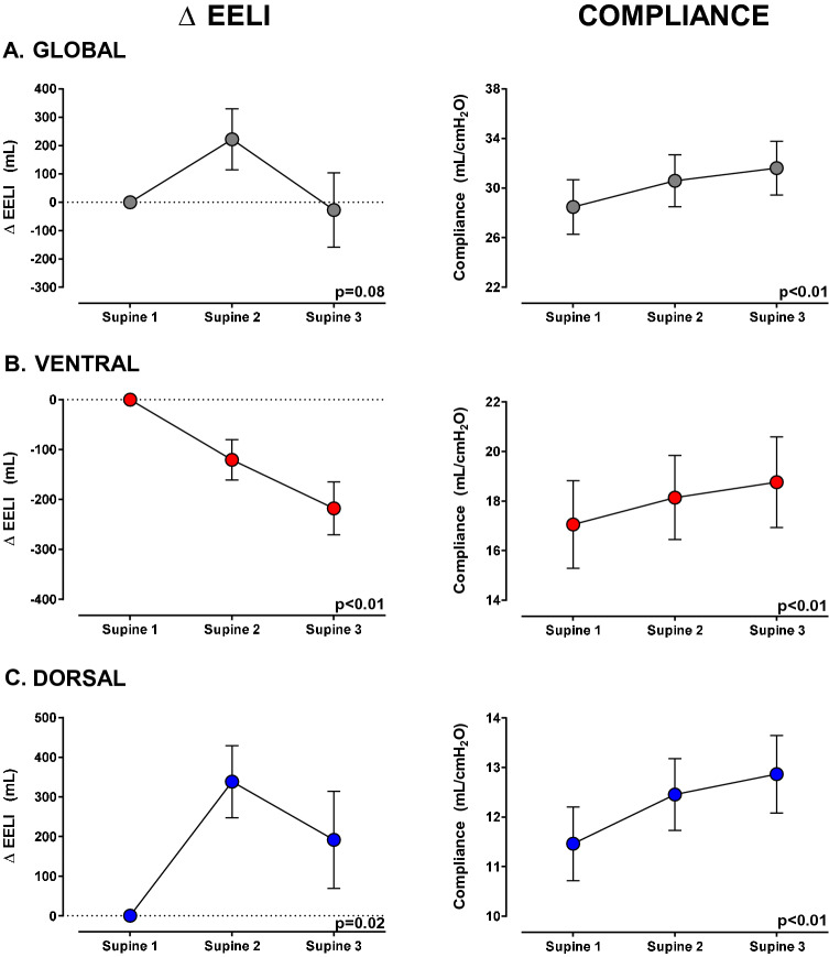
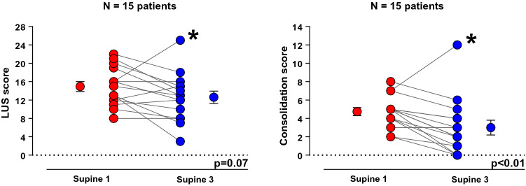
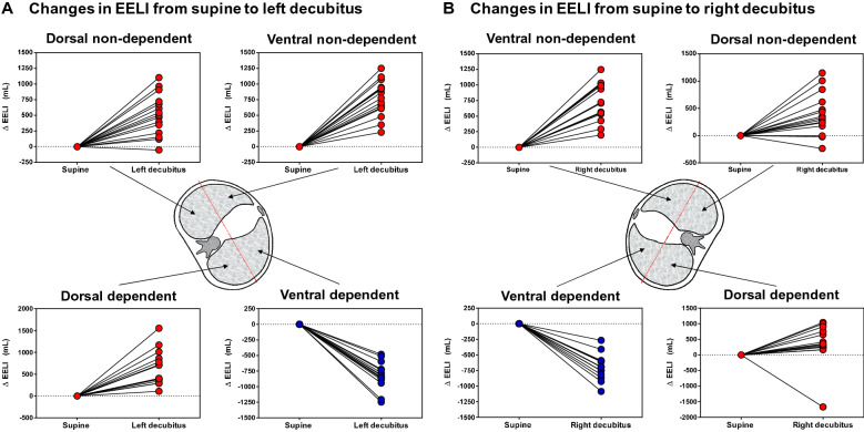
Results: Comparing supine baseline and final, respiratory compliance (29 ± 9 vs 32 ± 8 mL/cmH2O; p < 0.01) and PaO2/FIO2 ratio (138 ± 36 vs 164 ± 46 mmHg; p < 0.01) increased, while driving pressure (13 ± 2 vs 11 ± 2 cmH2O; p < 0.01) and lung ultrasound consolidation score decreased [5 (4-5) vs 2 (1-4); p < 0.01]. EELI decreased ventrally (218 ± 205 mL; p < 0.01) and increased dorsally (192 ± 475 mL; p = 0.02), while regional compliance increased in both ventral (11.5 ± 0.7 vs 12.9 ± 0.8 mL/cmH2O; p < 0.01) and dorsal regions (17.1 ± 1.8 vs 18.8 ± 1.8 mL/cmH2O; p < 0.01). Dorsal distribution of perfusion increased (64.8 ± 7.3% vs 66.3 ± 7.2%; p = 0.01).

Conclusions: Without increasing airway pressure, a sequential postural recruitment maneuver improves global and regional respiratory mechanics and gas exchange along with a redistribution of EELI from ventral to dorsal lung areas and less consolidation. Trial registration ClinicalTrials.gov, NCT04475068. Registered 17 July 2020, https://clinicaltrials.gov/ct2/show/NCT04475068.

Keywords: ARDS; COVID-19; PEEP; Postural lung recruitment.


Conflict of interest statement

The authors declare that they have no competing interests. LB’s laboratory has received research grants from Medtronic and Draeger and equipment from Sentec, Philps, Fisher Paykel and Air Liquide.

Figures

Fig. 1
Fig. 1
Protocol flowchart and EIT lung segmentation. The protocol flowchart (A) is shown at the top. The positioning sequence begins with the less ventilated lung evaluated by EIT positioned upwards in L1. In section B is shown the EIT lung segmentation by ROIs. To compare changes during supine position, the lungs were segmented into two equally sized ROIs: ventral and dorsal. To compare changes from supine to lateral position, the lungs positioned upwards (non-dependent) or downwards (dependent lung) during L1 or L2, were segmented into four ROIs or quadrants: ventral non-dependent, dorsal non-dependent, dorsal dependent, and ventral dependent. EIT: electrical impedance tomography; ROI: region of interest; L1: first lateral; L2: second lateral
Fig. 2
Fig. 2
End-expiratory lung impedance and compliance changes during supine position steps, from supine-1 (baseline) to supine-3 (after second lateral positioning), segmenting the lung into ventral and dorsal areas. The left column shows changes in the EELI, and the right column shows changes in compliance. The figures on the upper panel A show global lung changes, while the figures in the middle panel B show changes in the ventral (anterior half) part of the lung, and the figures on the bottom panel C show changes in the dorsal (posterior half) of the lung. The left column shows that global EELI did not change between supine-1 and supine-3, but the EELI decreased in the ventral region and increased in the dorsal region. This redistribution of EELI was accompanied by a progressive increase of the global and regional compliance (both ventral and dorsal) (right column). Δ EELI (mL): end-expiratory lung impedance change. Data are shown as mean ± SEM (standard error of mean). Mixed model was used for statistical analysis
Fig. 3
Fig. 3
Evaluation of aeration by lung ultrasound between supine-1 (baseline) and supine-3 (after second lateral positioning). The analysis of the individual data shows an improvement in LUS score (left figure) and consolidation score (right figure) in the majority of patients. A non-responder patient is identified with asterisk *LUS score: lung ultrasound score. Paired t test was used for the analysis of LUS score and Wilcoxon signed-rank test for the consolidation score
Fig. 4
Fig. 4
Changes in the end-expiratory lung impedance when going from supine to lateral position. Changes in end-expiratory lung impedance are presented, segmenting the lung into four quadrants from supine to left decubitus (left panel, A) and from supine to right decubitus (right panel, B). Regardless of the lateralized side, it is observed a decrease of EELI in the ventral quadrant of the lung placed down (dependent lung) and a significant increase in the other three quadrants. Δ EELI = End-expiratory lung impedance change. Data are shown as mean ± SEM

References

    1. Fan E, Del Sorbo L, Goligher EC, Hodgson CL, Munshi L, Walkey AJ, et al. An Official American Thoracic Society/European Society of Intensive Care Medicine/Society of Critical Care Medicine Clinical Practice Guideline: mechanical ventilation in adult patients with acute respiratory distress syndrome. Am J Respir Crit Care Med. 2017;195(9):1253–1263. doi: 10.1164/rccm.201703-0548ST. - DOI - PubMed
    1. Mora-Arteaga JA, Bernal-Ramirez OJ, Rodriguez SJ. The effects of prone position ventilation in patients with acute respiratory distress syndrome. A systematic review and meta analysis. Med Intensiva. 2015;39(6):359–372. doi: 10.1016/j.medin.2014.11.003. - DOI - PubMed
    1. Gonzalez-Seguel F, Pinto-Concha JJ, Aranis N, Leppe J. Adverse events of prone positioning in mechanically ventilated adults with ARDS. Respir Care. 2021 doi: 10.4187/respcare.09194. - DOI - PubMed
    1. Fan E, Wilcox ME, Brower RG, Stewart TE, Mehta S, Lapinsky SE, et al. Recruitment maneuvers for acute lung injury: a systematic review. Am J Respir Crit Care Med. 2008;178(11):1156–1163. doi: 10.1164/rccm.200802-335OC. - DOI - PubMed
    1. Constantin JM, Godet T, Jabaudon M, Bazin JE, Futier E. Recruitment maneuvers in acute respiratory distress syndrome. Ann Transl Med. 2017;5(14):290. doi: 10.21037/atm.2017.07.09. - DOI - PMC - PubMed

Associated data

LinkOut - more resources