Comparison of the vitality tests used in the dental clinical practice and histological analysis of the dental pulp
- PMID: 35150478
- PMCID: PMC9162753
- DOI: 10.17305/bjbms.2021.6841
Comparison of the vitality tests used in the dental clinical practice and histological analysis of the dental pulp
Abstract
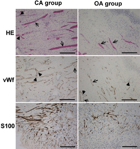
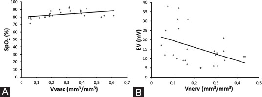
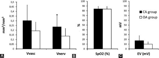
In dentistry, indirect diagnostic methods such as electrical sensibility testing and pulse oximetry are used to assess the status of the pulp. Our study aimed to determine the correlation between hemoglobin oxygen saturation and vascular volume density (Vvasc). We also wanted to examine an electrical sensibility test and the volume density of myelinated nerve fibers (Vnerv). Twenty-six intact permanent premolars were included in the study. For histological analysis, the pulp tissue was stained with hematoxylin-eosin and immunohistochemically for von Willebrand factor and S100 to detect blood vessels and myelinated nerve fibers, respectively. The stereological analysis was used to determine the Vvasc and Vnerv. Statistical analysis was done using the Pearson correlation test and Welch's ANOVA test. Histological analysis showed that the pulp tissue was strongly vascularized and innervated. A significant positive correlation was found between Vvasc and hemoglobin oxygen saturation levels (p=0.030). A significant negative correlation was found between Vnerv and the lowest electrical voltage that patient felt (p=0.033). According to the maturity of the dental apex, teeth were divided into a group with open (N=6, OA group) and closed apex (N=20, CA group). We found that pulps in the CA group had higher Vnerv than the OA group (p=0.037). In contrast, there were no significant differences in Vvasc of the pulp tissue (p=0.059), oxygen saturation (p=0.907), or electrical voltage (p=0.113) between both groups. We can conclude that the measurement of pulse oximetry and electrical sensibility test reflect the morphology of healthy pulp tissue independently of the maturity of the dental apex.
Conflict of interest statement
Conflicts of interest: The authors declare no conflicts of interest.
Figures
Similar articles
-
The use of pulse oximetry in evaluation of pulp vitality in immature permanent teeth.Dent Traumatol. 2016 Feb;32(1):43-7. doi: 10.1111/edt.12215. Epub 2015 Sep 10. Dent Traumatol. 2016. PMID: 26358664
-
Assessment of oxygen saturation in dental pulp of permanent teeth with periodontal disease.J Endod. 2014 Dec;40(12):1927-31. doi: 10.1016/j.joen.2014.08.009. Epub 2014 Oct 1. J Endod. 2014. PMID: 25282376
-
Oxygen saturation in the dental pulp of permanent teeth: a critical review.J Endod. 2014 Aug;40(8):1054-7. doi: 10.1016/j.joen.2014.04.011. Epub 2014 Jun 11. J Endod. 2014. PMID: 25069907 Review.
-
Oxygen saturation and perfusion index from pulse oximetry in adult volunteers with viable incisors.Acta Odontol Scand. 2016 Jul;74(5):411-5. doi: 10.3109/00016357.2016.1171898. Epub 2016 May 3. Acta Odontol Scand. 2016. PMID: 27140658
-
Pulp sensibility and vitality tests for diagnosing pulpal health in permanent teeth: a critical review.Int Endod J. 2017 Feb;50(2):135-142. doi: 10.1111/iej.12611. Epub 2016 Feb 11. Int Endod J. 2017. PMID: 26789282 Review.
Cited by
-
Expression of CD31, CD34, and smooth muscle actin (SMA) in endothelial cells of dental pulp vessels.Biomol Biomed. 2023 Dec 28;24(4):821-826. doi: 10.17305/bb.2023.9988. Biomol Biomed. 2023. PMID: 38153414 Free PMC article.
-
Assessment of dental pulp response to carries via MR T2mapping and histological analysis.BMC Oral Health. 2024 Apr 6;24(1):428. doi: 10.1186/s12903-024-04165-1. BMC Oral Health. 2024. PMID: 38582832 Free PMC article.
-
Dental Pulp Vascular Response to Early Stages of Caries.Int Dent J. 2024 Dec;74(6):1405-1412. doi: 10.1016/j.identj.2024.05.005. Epub 2024 Jun 8. Int Dent J. 2024. PMID: 38851930 Free PMC article.
-
The effect of orthodontic tooth movement on the sensitivity of dental pulp: A systematic review and meta-analysis.Heliyon. 2023 Mar 24;9(4):e14621. doi: 10.1016/j.heliyon.2023.e14621. eCollection 2023 Apr. Heliyon. 2023. PMID: 37025792 Free PMC article.
References
-
- Sigurdsson A. Pulpal diagnosis. Endod Top. 2003;5(1):12–25.
-
- Mejàre IA, Axelsson S, Davidson T, Frisk F, Hakeberg M, Kvist T, et al. Diagnosis of the condition of the dental pulp:A systematic review. Int Endod J. 2012;45(7):597–613. https://doi.org/10.1111/j.1365-2591.2012.02016.x. - PubMed
-
- Alghaithy RA, Qualtrough AJ. Pulp sensibility and vitality tests for diagnosing pulpal health in permanent teeth:A critical review. Int Endod J. 2017;50(2):135–42. https://doi.org/10.1111/iej.12611. - PubMed
-
- Abd-Elmeguid A, Yu DC. Dental pulp neurophysiology:Part 2. Current diagnostic tests to assess pulp vitality. J Can Dent Assoc. 2009;75(2):139–43. - PubMed
-
- Gopikrishna V, Pradeep G, Venkateshbabu N. Assessment of pulp vitality:A review. Int J Paediatr Dent. 2009;19(1):3–15. https://doi.org/10.1111/j.1365-263X.2008.00955.x. - PubMed
MeSH terms
Substances
LinkOut - more resources
Full Text Sources
Miscellaneous