. 2022 Mar;298(3):101712.
doi: 10.1016/j.jbc.2022.101712. Epub 2022 Feb 9.

The tocopherol transfer protein mediates vitamin E trafficking between cerebellar astrocytes and neurons

Affiliations

The tocopherol transfer protein mediates vitamin E trafficking between cerebellar astrocytes and neurons

L Ulatowski et al. J Biol Chem. 2022 Mar.

Abstract

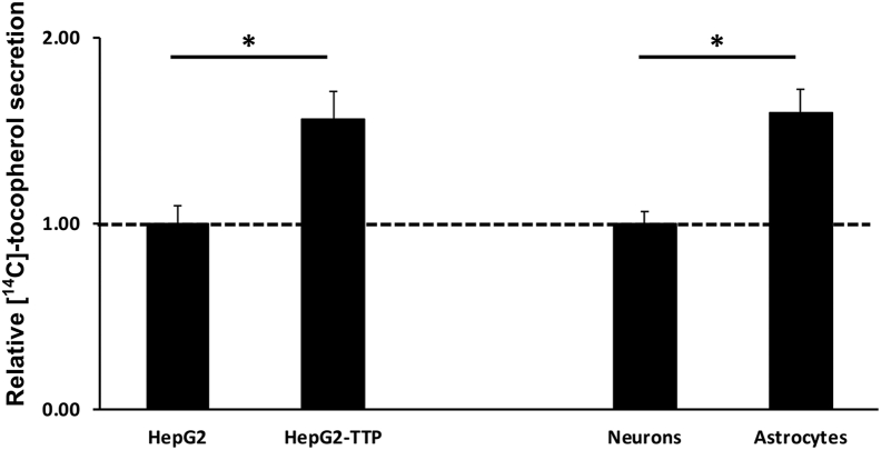
Alpha-tocopherol (vitamin E) is an essential nutrient that functions as a major lipid-soluble antioxidant in humans. The alpha-tocopherol transfer protein (TTP) binds α-tocopherol with high affinity and selectivity and regulates whole-body distribution of the vitamin. Heritable mutations in the TTPA gene result in familial vitamin E deficiency, elevated indices of oxidative stress, and progressive neurodegeneration that manifest primarily in spinocerebellar ataxia. Although the essential role of vitamin E in neurological health has been recognized for over 50 years, the mechanisms by which this essential nutrient is transported in the central nervous system are poorly understood. Here we found that, in the murine cerebellum, TTP is selectively expressed in glial fibrillary acidic protein-positive astrocytes, where it facilitates efflux of vitamin E to neighboring neurons. We also show that induction of oxidative stress enhances the transcription of the TtpA gene in cultured cerebellar astrocytes. Furthermore, secretion of vitamin E from astrocytes is mediated by an ABC-type transporter, and uptake of the vitamin into neurons involves the low-density lipoprotein receptor-related protein 1. Taken together, our data indicate that TTP-expressing astrocytes control the delivery of vitamin E from astrocytes to neurons, and that this process is homeostatically responsive to oxidative stress. These are the first observations that address the detailed molecular mechanisms of vitamin E transport in the central nervous system, and these results have important implications for understanding the molecular underpinnings of oxidative stress-related neurodegenerative diseases.

Keywords: lipid trafficking; lipid transport; tocopherol; tocopherol transfer protein; vitamin E.


Conflict of interest statement

Conflict of interest The authors declare that they have no conflicts of interest with the contents of this article.

Figures

Figure 1
Figure 1
Expression of TTP in the brain varies spatially and temporally and is restricted to astrocytes in the cerebellum.A, expression of TTP mRNA in the mouse brain. Tissue lysates from indicated brain regions of 10-week-old C57BL6J WT mice were used to determine mRNA levels of TtpA by RT–PCR. B, expression of TTP protein in the mouse brain. Tissue lysates from indicated brain regions of 10-week-old C57BL6J WT mice (except where indicated by asterisk; 8 days old) were analyzed by anti-TTP immunoblotting. Two hundred micrograms of protein of the indicated tissues were used. Heart extract is shown as a negative control. Mice were 10 weeks old (except the cerebellum samples, which were prepared from 8-day-old mice). C, anti-TTP immunohistochemistry staining of paraffin-embedded C57BL6J WT (left) and Ttpa−/− (right) mouse cerebella sections counterstained with hematoxylin. The black arrows demarcate the positive TTP signal, whereas the bold white arrows point to the Purkinje neuron soma. The scale bar represents 20 μm. B, cerebellum; BS, brainstem; CX, cerebellar cortex; GL, granule cell layer; HP, hippocampus; MB, midbrain; ML, molecular layer; PCL, Purkinje cell layer; TTP, alpha-tocopherol transfer protein; WB, whole brain.
Figure 2
Figure 2
Ttpa is preferentially expressed in cerebellar astrocytes. Primary dissociated cerebella cells harvested from 8-day-old C57BL6J WT mice were maintained in culture as described in the Experimental procedures section for 2 days (A) and 8 days (B) prior to immunostaining with the neuronal marker β-tubulin III (green) and the astrocyte-specific marker GFAP (red). The scale bars represent 20 μm. C, expression of TTP mRNA was measured in astrocyte-enriched or neuron-enriched cerebellar cultures using real-time RT–PCR. Expression was normalized to 18 s. Error bars indicate standard deviations, and asterisk denotes p > 0.05. GFAP, glial fibrillary acidic protein; TTP, alpha-tocopherol transfer protein.
Figure 3
Figure 3
TTP is expressed in astrocytes. Primary dissociated astrocyte-enriched (A) and neuron-enriched (B) cerebellar cultures and cerebellar organotypic slice cultures (C and D) harvested from an 8-day-old C57BL6J WT mice were maintained in culture prior to immunofluorescence with antibodies against TTP (A and B; green or C and D; red) and the astrocyte marker GFAP (A and C; red) or the neuronal marker β-tubulin III (B and D; red). Note the yellow in the merged images of (A and C) indicates colocalization. DAPI (blue) staining identifies the nuclei. The scale bars represent 20 μm. DAPI, 4′,6-diamidino-2-phenylindole; GFAP, glial fibrillary acidic protein; TTP, alpha-tocopherol transfer protein.
Figure 4
Figure 4
Vitamin E preferentially localizes to GFAP-positive and TTP-expressing cerebellar astrocytes. Merged images of primary dissociated cerebella cultures harvested from 8-day-old WT mice maintained in culture for 6 days prior to “loading” with NBD-tocopherol (green) and coimmunostaining with the astrocyte marker GFAP (red; A), the neuronal marker β-tubulin III (red; B), or TTP (red; C). White arrows in inset images indicate the NBD-tocopherol colocalization. The scale bars represent 20 μm. GFAP, glial fibrillary acidic protein; NBD, nitrobenzoxadiazole, TTP, alpha-tocopherol transfer protein.
Figure 5
Figure 5
Vitamin E localizes to lysosomes and mitochondria in primary neurons and astrocytes and is not affected by the presence of TTP. Merged images of Ttpa+/+ primary dissociated neuron (A and B) and astrocyte (C and D) cerebella cells maintained in culture for 24 h and 7 days, respectively, from Ttpa+/+ (A, C, E, and G) and Ttpa−/− (B, D, F, and H) mice prior to an overnight loading with BODIPY-tocopherol (green) and a subsequent staining with LysoTracker Red (A and C; red) to mark the lysosomes or with MitoTracker Red (B and D; red) to visualize mitochondria. The yellow in the perinuclear region of the neurons (A) and astrocytes (C) indicates localization of BODIPY-tocopherol to lysosomes. The yellow in the overlaid images indicates that BODIPY-tocopherol localizes to mitochondria in the astrocytes (D) but not in the neurons. These findings were unchanged in Ttpa−/− mice (B, D, F, and H), indicating that this distribution pattern is not affected by the presence of TTP. The scale bars represent 10 μm. TTP, alpha-tocopherol transfer protein.
Figure 6
Figure 6
Secretion of vitamin E is enhanced in TTP-expressing hepatocytes and astrocytes. HepG2-TetOn-TTP cells (left panel) were induced to express TTP with doxycycline (1 μg/ml) where indicated. Primary cerebellar neurons and astrocytes (right panel) were harvested from TtpA+/+ mice and cultured as described in the Experimental procedures section. Cells were loaded for 48 h with [14C]-α-tocopherol, followed by washes, and a secretion period of 24 h. Asterisks denote significant difference in secretion between the neurons and astrocytes or the induced and uninduced HepG2 cells (p < 0.05; Student's t test). TTP, alpha-tocopherol transfer protein.
Figure 7
Figure 7
Expression of the TtpA gene in cerebellar astrocytes is regulated by oxidative stress. Real-time PCR results of normalized Ttpa expression (black bars; left axis) and H2O2-induced oxidative stress, as determined by the DCF-DA assay (gray bars; right axis), in primary astrocytes (A) or neurons (B). Where indicated, tocopherol was added 16 h prior to H2O2 addition. Asterisks indicate significance of p < 0.05 compared with nontreated cells (one-way ANOVA with Tukey's post hoc testing). DCF-DA, dichlorodihydrofluorescein diacetate.
Figure 8
Figure 8
Working model of vitamin E transport in the cerebellum. Alpha-tocopherol accumulates in astrocytes, such as the cerebellar Bergman glia cells, where it is stored in endocytic vesicles. Astrocyte-expressed TTP facilitates the secretion of vitamin E via an ABC transporter to the extracellular milieu, where the vitamin is assembled into ApoE-containing lipoprotein particles, which are taken up by neighboring neurons (e.g., cerebellar Purkinje cells) via LRP1. Elevation of oxidative stress enhances expression of TTP in astrocytes, thereby facilitating egress of the vitamin to neurons, which are especially vulnerable to oxidative stress. Loss-of-function mutations in TTP, such as those present in heritable AVED cases, impair vitamin E trafficking, thereby eliciting oxidative injury to neurons and giving rise to cerebellar ataxia. ApoE, apolipoprotein E; AVED, ataxia with vitamin E deficiency; LRP1, low-density lipoprotein receptor–related protein 1; TTP, alpha-tocopherol transfer protein.

References

    1. Ulatowski L., Manor D. Vitamin E trafficking in neurologic health and disease. Annu. Rev. Nutr. 2013;33:87–103. - PMC - PubMed
    1. Burton G.W., Cheeseman K.H., Doba T., Ingold K.U., Slater T.F. Vitamin E as an antioxidant in vitro and in vivo. Ciba Found. Symp. 1983;101:4–18. - PubMed
    1. Ingold K.U., Webb A.C., Witter D., Burton G.W., Metcalfe T.A., Muller D.P. Vitamin E remains the major lipid-soluble, chain-breaking antioxidant in human plasma even in individuals suffering severe vitamin E deficiency. Arch. Biochem. Biophys. 1987;259:224–225. - PubMed
    1. Muller D.P., Lloyd J.K. Effect of large oral doses of vitamin E on the neurological sequelae of patients with abetalipoproteinemia. Ann. N. Y. Acad. Sci. 1982;393:133–144. - PubMed
    1. Runge P., Muller D.P., McAllister J., Calver D., Lloyd J.K., Taylor D. Oral vitamin E supplements can prevent the retinopathy of abetalipoproteinaemia. Br. J. Ophthalmol. 1986;70:166–173. - PMC - PubMed