. 2022 Mar;298(3):101713.
doi: 10.1016/j.jbc.2022.101713. Epub 2022 Feb 9.

The tetrameric structure of Plasmodium falciparum phosphoglycerate mutase is critical for optimal enzymatic activity

Affiliations

The tetrameric structure of Plasmodium falciparum phosphoglycerate mutase is critical for optimal enzymatic activity

Ankita Tehlan et al. J Biol Chem. 2022 Mar.

Abstract

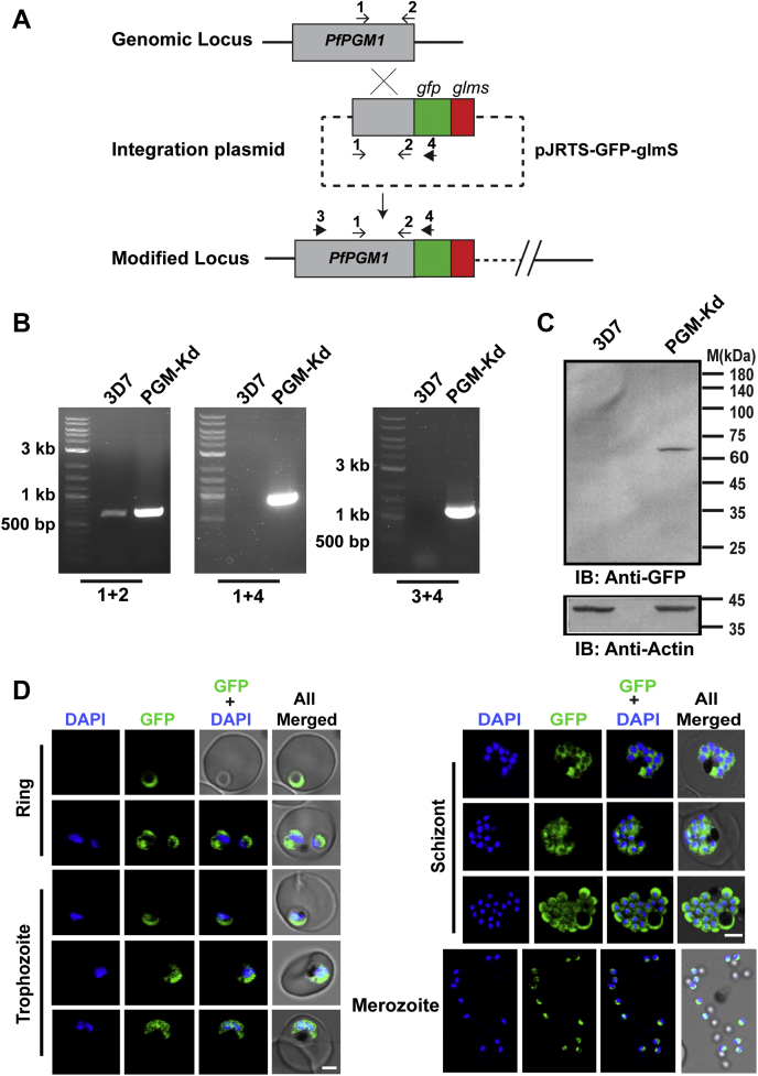
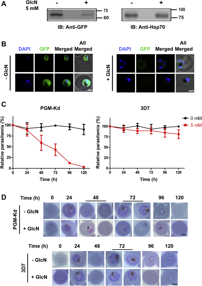
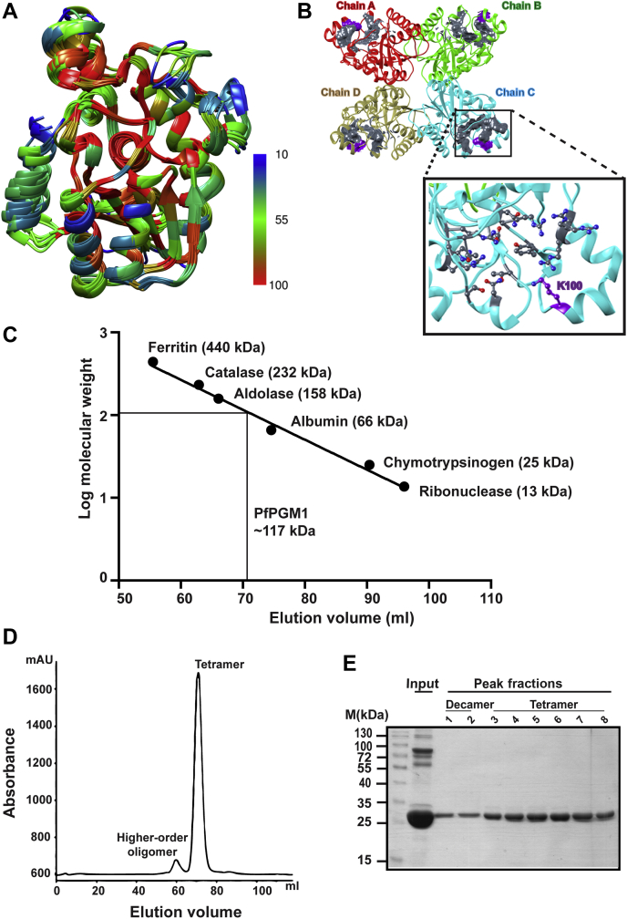
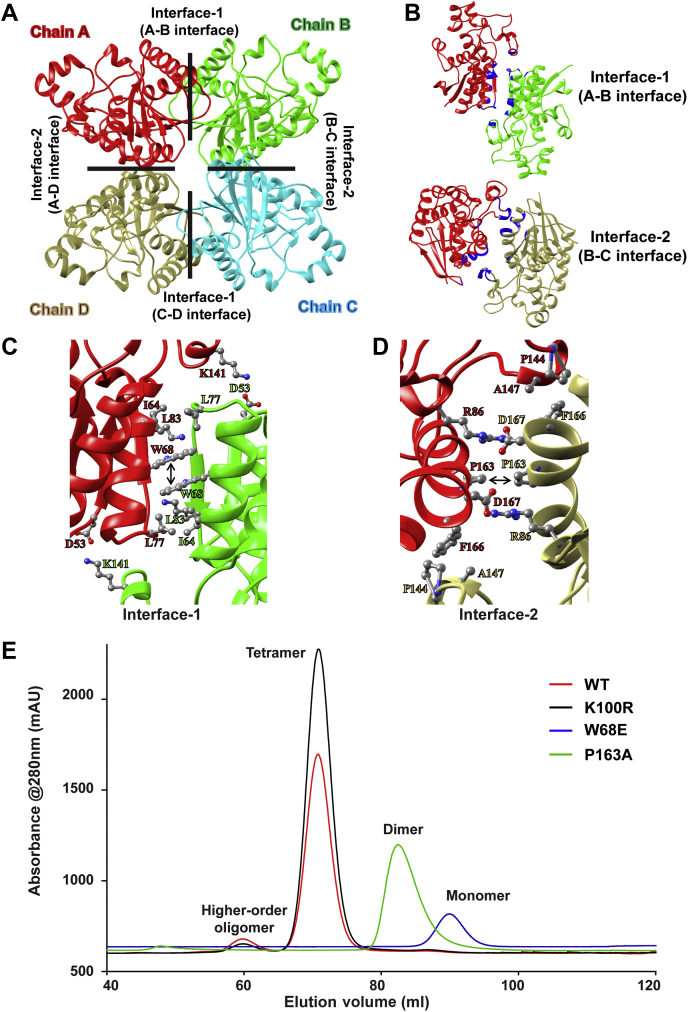
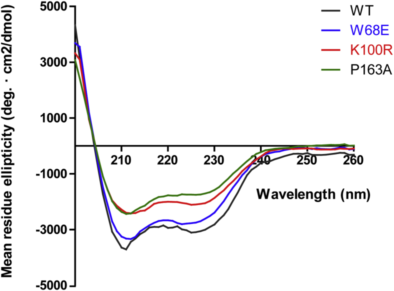
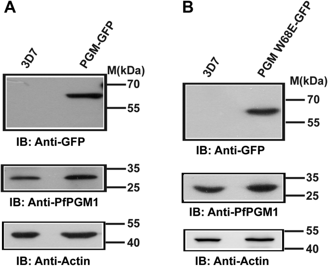
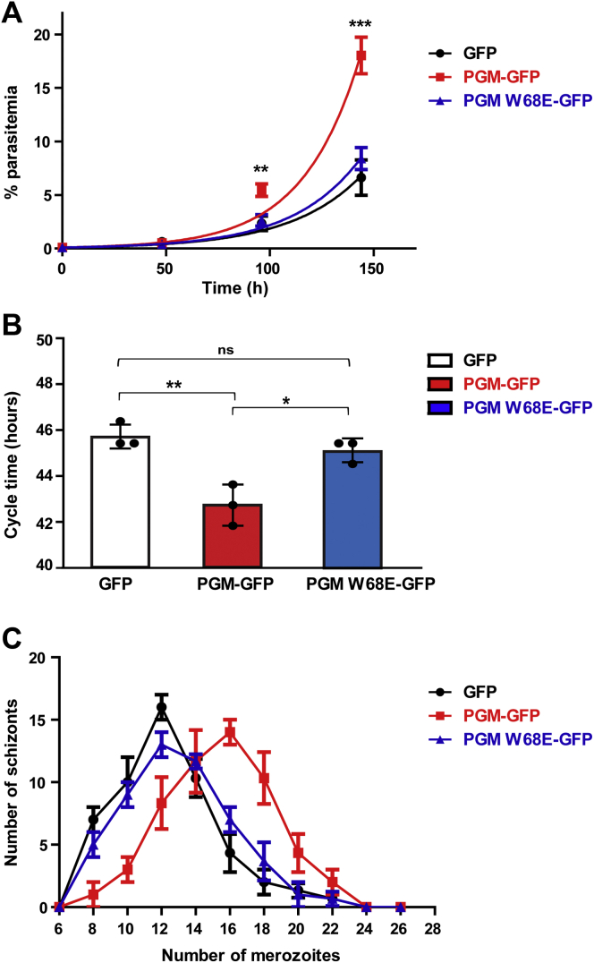
The glycolytic enzyme phosphoglycerate mutase (PGM) is of utmost importance for overall cellular metabolism and has emerged as a novel therapeutic target in cancer cells. This enzyme is also conserved in the rapidly proliferating malarial parasite Plasmodium falciparum, which have a similar metabolic framework as cancer cells and rely on glycolysis as the sole energy-yielding process during intraerythrocytic development. There is no redundancy among the annotated PGM enzymes in Plasmodium, and PfPGM1 is absolutely required for the parasite survival as evidenced by conditional knockdown in our study. A detailed comparison of PfPGM1 with its counterparts followed by in-depth structure-function analysis revealed unique attributes of this parasitic protein. Here, we report for the first time the importance of oligomerization for the optimal functioning of the enzyme in vivo, as earlier studies in eukaryotes only focused on the effects in vitro. We show that single point mutation of the amino acid residue W68 led to complete loss of tetramerization and diminished catalytic activity in vitro. Additionally, ectopic expression of the WT PfPGM1 protein enhanced parasite growth, whereas the monomeric form of PfPGM1 failed to provide growth advantage. Furthermore, mutation of the evolutionarily conserved residue K100 led to a drastic reduction in enzymatic activity. The indispensable nature of this parasite enzyme highlights the potential of PfPGM1 as a therapeutic target against malaria, and targeting the interfacial residues critical for oligomerization can serve as a focal point for promising drug development strategies that may not be restricted to malaria only.

Keywords: Plasmodium falciparum; enzyme kinetics; glmS; knockdown; phosphoglycerate mutase; tetramer.


Conflict of interest statement

Conflict of interest The authors declare that they have no conflict of interest with the contents of this article.

Figures

Figure 1
Figure 1
PfPGM1 is expressed throughout the parasite developmental stages.A, the integration strategy schematic for endogenous tagging of PfPGM1 at the genomic locus by single crossover homologous recombination. The primers used in cloning (1, 2) and the diagnostic PCR (3, 4) are indicated by arrows and numbered accordingly. GFP and glmS tag are represented by green and red boxes, respectively. B, diagnostic PCR with the indicated primer sets to confirm integration at the PfPGM1 genomic locus in the obtained transgenic parasites (denoted PGM-Kd). 3D7 parasites, having the unmodified genomic locus, were used as control. C, immunoblotting using antibodies against GFP confirms the expression of GFP-fusion protein in the PGM-Kd parasite line. PfActin served as a loading control. D, live cell microscopy images demonstrating the abundance and subcellular localization of the GFP-tagged PfPGM1 protein (green) in the PGM-Kd parasites. 4′,6-diamidino-2-phenylindole (DAPI) was used to stain the nuclei. The scale bar represents 2 μm. The sequences of the primers (1–4) are listed in the Table S3 (S.No. 11–14). Kd, knockdown; PGM, phosphoglycerate mutase.
Figure 2
Figure 2
PfPGM1 is indispensable for the parasite growth and development.A, Western blot analysis using antibodies against GFP illustrating the regulation of PfPGM1 expression via glmS riboswitch. The PfPGM1 expression levels were downregulated following 24 h of glucosamine treatment (5 mM) in synchronized ring parasites. PfHsp70 was used as a loading control. B, confocal microscopy live cell images illustrating the depletion of PfPGM1 and its effect on parasite phenotype. The scale bar denotes 2 μm. C, parasite survival curves demonstrating relative parasitemia in the absence and presence of 5 mM glucosamine in PGM-Kd (left) and 3D7 parasites taken as control (right). The graphs represent mean ± SD of at least three independent experiments. D, Giemsa-stained erythrocyte smears depicting the progression of the parasite in the presence and absence of PfPGM1 and the effect of PfPGM1 knockdown on parasite phenotype (top panel). Effect of 5 mM glucosamine on the phenotype of 3D7 parasites, taken as control (bottom panel). The scale bar denotes 2 μm. Kd, knockdown; PGM, phosphoglycerate mutase.
Figure 3
Figure 3
PfPGM1 structure and its oligomeric status.A, structural comparison of PfPGM1 and its homologs. Monomeric subunits of each homolog were structurally aligned to PfPGM1 structure, and the corresponding RMSD values are provided in Table S1. Amino acid residues are colored as per the color code. Blue and red colors denote the least and maximum conservation, respectively at the corresponding amino acid position among the homologs. B, the tetrameric structure of PfPGM1 illustrating the active site in gray surface view and labeled. The position of K100 is shown in the inset in purple. C, standard curve of known molecular mass proteins separated with size-exclusion chromatography. The solid line interpolation corresponds to the tetrameric form of PfPGM1. D, size-exclusion chromatogram of PfPGM1 displaying elution peaks of the two oligomeric forms. E, SDS-PAGE of the peak fractions in comparison with input highlighting the purity of the eluted fractions. PGM, phosphoglycerate mutase.
Figure 4
Figure 4
Key amino acid residues involved in the oligomerization of PfPGM1.A, the four subunits of PfPGM1 are represented as Chains A to D. The interface-1 formed between chains A-B and C-D and the interface-2 formed between chains B-C and A-D are marked by black lines and labeled accordingly. B, depicts interface-1 (chain A-B interface) on top and interface-2 (chain A-D interface) at the bottom. The interface residues are highlighted in blue color. Amino acid residues at PfPGM1 oligomerization interface-1 and interface-2 are shown in (C) and (D) respectively. The residues forming the hydrophobic interactions (W68-W68 at interface-1 and P163-P163 at interface-2) in the center of both the interfaces is marked by the black arrow. The interaction profile at the respective interfaces is shown in Fig. S4 in more detail. E, size-exclusion chromatogram demonstrating the elution peaks and oligomeric forms of WT protein and mutants. PGM, phosphoglycerate mutase.
Figure 5
Figure 5
Comparison of Far-UV CD spectra of PfPGM1 mutant proteins with the WT protein. The graph is representative of data of one experiment from two independent repeats. PGM, phosphoglycerate mutase.
Figure 6
Figure 6
Ectopic expression of PfPGM1-GFP and PfPGM1 W68E-GFP. Immunoblotting with antibodies against GFP and PfPGM1 in the transgenic parasites expressing, (A) PfPGM1-GFP and (B) PfPGM1 W68E-GFP, illustrating the expression profile of GFP-fusion protein (top panel) and the endogenous protein (middle panel). PfActin served as a loading control (bottom panel). 3D7 parasites were taken as control. PGM, phosphoglycerate mutase.
Figure 7
Figure 7
PfPGM1 enhances parasite growth and proliferation rate.A, the growth of transgenic parasite lines expressing the WT tetrameric protein, monomer mutant W68E, and GFP only were monitored for three cycles. B, duration of IDC for the three parasite lines. C, number of merozoites produced in different schizonts in the three transgenic parasite lines. The graphs represent mean ± SD of three independent experiments. Statistical significance of the data is represented as: ns p > 0.05, ∗ p ≤ 0.05, ∗∗ p ≤ 0.01 and ∗∗∗ p ≤ 0.001. IDC, intra-erythrocytic developmental cycle; PGM, phosphoglycerate mutase.

References

    1. World Health Organization . World Health Organization; Geneva: 2020. World Malaria Report 2020: 20 Years of Global Progress and Challenges.
    1. Maier A.G., Matuschewski K., Zhang M., Rug M. Plasmodium falciparum. Trends Parasitol. 2019;35:481–482. - PubMed
    1. Roth E., Jr. Plasmodium falciparum carbohydrate metabolism: A connection between host cell and parasite. Blood Cells. 1990;16:453–460. - PubMed
    1. Mehta M., Sonawat H.M., Sharma S. Glycolysis in Plasmodium falciparum results in modulation of host enzyme activities. J. Vector Borne Dis. 2006;43:95–103. - PubMed
    1. Alam A., Neyaz M.K., Ikramul Hasan S. Exploiting unique structural and functional properties of malarial glycolytic enzymes for antimalarial drug development. Malar. Res. Treat. 2014;2014:451065. - PMC - PubMed

MeSH terms

Substances

LinkOut - more resources