Review
. 2022 Feb 12;12(1):5.
doi: 10.1186/s13395-022-00289-6.

Muscle stem cell adaptations to cellular and environmental stress

Affiliations
Review

Muscle stem cell adaptations to cellular and environmental stress

Maria Vittoria Gugliuzza et al. Skelet Muscle. .

Abstract

Background: Lifelong regeneration of the skeletal muscle is dependent on a rare population of resident skeletal muscle stem cells, also named 'satellite cells' for their anatomical position on the outside of the myofibre and underneath the basal lamina. Muscle stem cells maintain prolonged quiescence, but activate the myogenic programme and the cell cycle in response to injury to expand a population of myogenic progenitors required to regenerate muscle. The skeletal muscle does not regenerate in the absence of muscle stem cells.

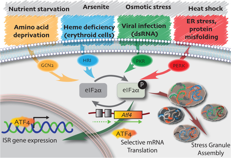
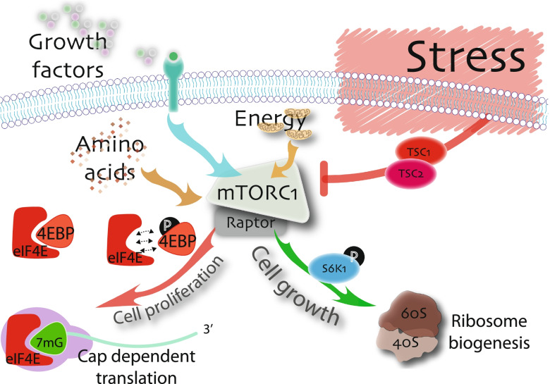
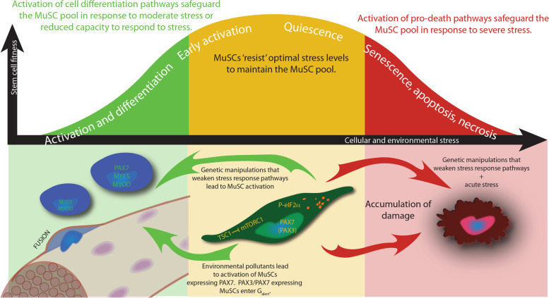
Main body: The notion that lifelong regeneration of the muscle is dependent on a rare, non-redundant population of stem cells seems contradictory to accumulating evidence that muscle stem cells have activated multiple stress response pathways. For example, muscle stem cell quiescence is mediated in part by the eIF2α arm of the integrated stress response and by negative regulators of mTORC1, two translational control pathways that downregulate protein synthesis in response to stress. Muscle stem cells also activate pathways to protect against DNA damage, heat shock, and environmental stress. Here, we review accumulating evidence that muscle stem cells encounter stress during their prolonged quiescence and their activation. While stress response pathways are classically described to be bimodal whereby a threshold dictates cell survival versus cell death responses to stress, we review evidence that muscle stem cells additionally respond to stress by spontaneous activation and fusion to myofibres.

Conclusion: We propose a cellular stress test model whereby the prolonged state of quiescence and the microenvironment serve as selective pressures to maintain muscle stem cell fitness, to safeguard the lifelong regeneration of the muscle. Fit muscle stem cells that maintain robust stress responses are permitted to maintain the muscle stem cell pool. Unfit muscle stem cells are depleted from the pool first by spontaneous activation, or in the case of severe stress, by activating cell death or senescence pathways.

Keywords: MuSC; Muscle stem cell; Stress response pathways; Translational control of gene expression.


Conflict of interest statement

The authors declare that they have no competing interests.

Figures

Fig. 1
Fig. 1
Living outside the comfort zone: quiescent MuSCs adapt to cellular and environmental stress. A stylised MuSC (green) highlighted over the skeletal muscle myofibres (grey). MuSCs maintain low levels of protein synthesis by phosphorylation of eIF2α and TSC1 inhibition of mTORC1 activity. Genetic inactivation of these stress response pathways in MuSCs leads to Galert or spontaneous activation. P-eIF2α leads to the assembly of DDX6(+) RNA granules (orange) in MuSCs, which resemble stress granules and may functionally serve as sights of mRNA sequestration. P-eIF2α also leads to translational reprogramming of mRNAs that confer stem cell properties on quiescent, self-renewing, or expanding MuSCs. In the nucleus of quiescent MuSCs (light green), elevated DNA-PKcs levels ensure efficient and accurate DNA repair. Expression of the paired homeodomain transcription factor Pax3 marks a subset of MuSCs with increased resistance to multiple stresses, including DNA damage and environmental pollutants. MuSCs that do not express Pax3 spontaneously activate when challenged with the environmental pollutant TCDD. Quiescent MuSCs express mRNAs for heat shock proteins Hsp40, Hsp70, and Hsp90 (brown) and upregulate these chaperones during early activation. These chaperones may be required to counteract the accumulation of misfolded proteins
Fig. 2
Fig. 2
The integrated stress response. In response to various sources of cellular and environmental stress, a family of four eIF2α kinases phosphorylate (black P) eIF2α (light grey). GCN2 (yellow) responds to amino acid deficiency, HRI (blue) responds to heme deficiency in erythroid cells, and PKR (green) responds to the presence of double-stranded RNA coincident with viral infection and PERK (red) responds to endoplasmic reticulum stress. Additional stresses that phosphorylate eIF2α, for which the corresponding kinase is unknown, are indicated (grey). Phosphorylated eIF2α leads to a global repression of translation. Accumulation of a pool of mRNAs, stalled at the initiation step of translation, seed the assembly of stress granules (maroon arrow). In contrast P-eIF2α reprograms translation to favour a subset of mRNAs, such as those for activating transcription factor Atf4 (orange), that contain uORFs in their 5’UTR. P-eIF2α-dependent translation of Atf4 and subsequent nuclear localisation of ATF4 protein initiate the integrated stress response (dark green arrow), a pro-survival pathway
Fig. 3
Fig. 3
The mTORC1 pathway in stress. The presence of growth factors (grey, green, purple circles), abundant amino acids (brown diamonds), and cellular energy (light brown mitochondria) positively regulated mTORC1 to regulate cell growth pathways through the phosphorylation of S6K1 kinase (green arrow), and cell proliferation pathways (red arrow) through the phosphorylation of eIF4E binding protein (4E-BP). Phosphorylated 4E-BP no longer competes for eIF4E binding, permitted eIF4E to initiate cap-dependent translation. eIF4E is the cap binding protein that functions within the eIF4F tertiary complex (purple) along with eIF4G and eIF4A (not shown). Cellular and environmental stress activates the TSC1/TSC2 complex to inhibit mTORC1 signalling, leading to the repression of these pathways, with consequent decrease in cell growth and proliferation
Fig. 4
Fig. 4
MuSC fitness in relationship to cellular and environmental stress. Cell fates associated with increasing stress are indicated along the modified Yerkes-Dodson curve (white text). (Yellow zone) Optimal levels of cellular and environmental stress are required to maintain the quiescent MuSC pool (dark green cell). Activation of stress response pathways that inhibit protein synthesis, including phosphorylation of eIF2α (P-eIF2α) and TSC1 inhibition of mTORC1 signalling, are required for MuSC quiescence and self-renewal. In addition, a subset of quiescent MuSCs expressing PAX3 (PAX3) exhibit enhanced resistance to stress. Activation of stress response pathways in quiescent MuSCs are also illustrated by the presence of P-eIF2α-dependent RNA granules (orange foci). (Green zone) MuSCs with reduced cellular fitness are removed from the MuSC pool by spontaneous activation (blue cells) and contribution to the myofibre (fusing blue cell with the brown myofibre; myonuclei are indicated in purple). Genetic inactivation of P-eIF2α leads to activation and differentiation of MuSCs. Genetic inactivation of Tsc1 and exposure to the environmental pollutant TCDD leads to the Galert state of early activation and/or full MuSC activation and differentiation. (Red zone) MuSCs that encounter severe stress, for example accumulating damage associated with aging, or proliferative stress associated with chronic muscle degeneration, are removed from the stem cell pool by the activation of cell senescence or death pathways (bloated red cell)

References

    1. Mauro A. Satellite cell of skeletal muscle fibers. J Biophys Biochem Cytol. 1961;9:493–495. - PMC - PubMed
    1. Crist CG, Montarras D, Buckingham M. Muscle satellite cells are primed for myogenesis but maintain quiescence with sequestration of Myf5 mRNA targeted by microRNA-31 in mRNP granules. Cell Stem Cell. 2012;11(1):118–126. - PubMed
    1. Fujita R, Zismanov V, Jacob J-M, Jamet S, Asiev K, Crist C. Fragile X mental retardation protein regulates skeletal muscle stem cell activity by regulating the stability of Myf5 mRNA. Skeletal Muscle. 2017;7(1):18. - PMC - PubMed
    1. Hausburg MA, Doles JD, Clement SL, Cadwallader AB, Hall MN, Blackshear PJ, et al. Post-transcriptional regulation of satellite cell quiescence by TTP-mediated mRNA decay. Elife. 2015;4:e03390. - PMC - PubMed
    1. De Morree A, van Velthoven CTJ, Gan Q, Salvi JS, Klein JDD, Akimenko I, et al. Staufen1 inhibits MyoD translation to actively maintain muscle stem cell quiescence. Proc Natl Acad Sci USA. 2017;114(43):E8996–E9005. - PMC - PubMed

Publication types

MeSH terms

Grants and funding

LinkOut - more resources