. 2022 Mar;298(3):101716.
doi: 10.1016/j.jbc.2022.101716. Epub 2022 Feb 11.

A mouse model of inherited choline kinase β-deficiency presents with specific cardiac abnormalities and a predisposition to arrhythmia

Affiliations

A mouse model of inherited choline kinase β-deficiency presents with specific cardiac abnormalities and a predisposition to arrhythmia

Mahtab Tavasoli et al. J Biol Chem. 2022 Mar.

Abstract

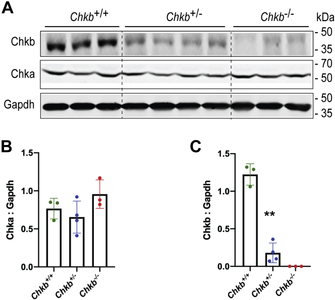
The CHKB gene encodes choline kinase β, which catalyzes the first step in the biosynthetic pathway for the major phospholipid phosphatidylcholine. Homozygous loss-of-function variants in human CHKB are associated with a congenital muscular dystrophy. Dilated cardiomyopathy is present in some CHKB patients and can cause heart failure and death. Mechanisms underlying a cardiac phenotype due to decreased CHKB levels are not well characterized. We determined that there is cardiac hypertrophy in Chkb-/- mice along with a decrease in left ventricle size, internal diameter, and stroke volume compared with wildtype and Chkb+/- mice. Unlike wildtype mice, 60% of the Chkb+/- and all Chkb-/- mice tested displayed arrhythmic events when challenged with isoproterenol. Lipidomic analysis revealed that the major change in lipid level in Chkb+/- and Chkb-/- hearts was an increase in the arrhythmogenic lipid acylcarnitine. An increase in acylcarnitine level is also associated with a defect in the ability of mitochondria to use fatty acids for energy and we observed that mitochondria from Chkb-/- hearts had abnormal cristae and inefficient electron transport chain activity. Atrial natriuretic peptide (ANP) is a hormone produced by the heart that protects against the development of heart failure including ventricular conduction defects. We determined that there was a decrease in expression of ANP, its receptor NPRA, as well as ventricular conduction system markers in Chkb+/- and Chkb-/- mice.

Keywords: acylcarnitine; cardiac muscle; heart disease; lipid; metabolism; muscular dystrophy; phosphatidylcholine.


Conflict of interest statement

Conflict of interest The authors declare that they have no conflicts of interest with the contents of this article.

Figures

Figure 1
Figure 1
Chka protein levels in heart samples from Chkb-deficient mice.A, Western blot of heart samples from three distinct (lanes 1–3) Chkb+/+, four distinct (lanes 4–7) Chkb+/, and three distinct (lanes 8–10) Chkb/ mice probed with anti-Chkb, anti-Chka, and anti-Gapdh antibodies. B and C, densitometry of the Western blot data. Loss of Chkb does not affect Chka level in heart. Values are means ± SD; data were analyzed using one-way ANOVA with Tukey’s multiple comparison test. ∗∗p< 0.01 (n = 3–4 females per group).
Figure 2
Figure 2
Chkb deficiency results in defects in heart function.A, body weight was recorded for 5-month-old Chkb+/+ (five male, four female), Chkb+/− (four male, eight female), and Chkb−/− (three male, three female) mice. Hearts were surgically removed from anesthetized mice and trimmed of extracardiac tissue. Heart weight was measured (B) and heart weight to body weight ratio (heart weight/body weight), an index of hypertrophy, was calculated (C). Values are presented as mean ± SD. Significance was calculated using one-way ANOVA with Tukey’s multiple comparison test for each specific time point. ∗∗p < 0.01 (n = 6–12 mice/group). D, representative M-mode images of 20-week-old Chkb+/+, Chkb+/−, and Chkb−/− mice. E, left ventricle mass; LV Mass AW (mg). F, left ventricle internal diameter during systole; LVID;s (mm). G, left ventricle internal diameter during diastole; LVID;d (mm). H, ejection fraction (EF, %). I, stroke volume (μl) and (J) Cardiac output (ml/min). Data were analyzed using one-way ANOVA (p < 0.05) with Tukey’s multiple comparison test for each specific time point; ∗p < 0.05, ∗∗p < 0.01 (n = 3–12 mice/group). ns, not significant.
Figure 3
Figure 3
Increased arrhythmic events due to Chkb deficiency.A, lead II ECG trace pattern of Chkb+/+ mice showing regular ECG pattern with defined P, QRS, and T waves at baseline and after isoproterenol (ISO). Chkb+/ and Chkb−/− mice showing regular ECG pattern at baseline but display arrhythmic events when challenged with ISO (highlighted with red circles). Quantification of arrhythmic events over 15 min (B) RR intervals (C), PR interval (D), heart rate (bpm) (E), ST height (mV) (F), QTc (s) (G), and QRS intervals (s) (H) at baseline and after treatment with ISO. Each bar represents mean ± SD. Data were analyzed using one-way ANOVA with Tukey’s multiple comparisons post hoc test; ∗∗p< 0.01 (n = 5–9 mice per group).
Figure 4
Figure 4
Chkb deficiency alters the cardiac lipid profile of Chkb+/and Chkb/mice and decreases mitochondrial β-oxidation capacity.A, PC synthesis is integrated with the synthesis of other major phospholipid classes, as well as AcCa, fatty acids, and the neutral lipids diacylglycerol (DG) and triacylglycerol (TG). B, comparison of expression levels of major glycerophospholipids (PC, PE, PI, PG, PS, CL, DG, and TG) and their metabolites (LPC, LPE) as well as sphingomyelin (SM), ceramide (Cer), and acyl carnitine (AcCa) between Chkb+/+, Chkb+/, and Chkb/ mice (n = 3 mice per group). The analysis was performed on 5-month-old heart samples. Each dot represents an individual fatty acyl lipid species, and the bar represents total mass. All statistical comparisons were performed pair-wise and log scaled. That is, a fold-change was calculated between the Chkb+/ and Chkb/ mice and controls for each unique lipid species, and a nonparametric statistical test was used to assess if the fold-changes were statistically significant from 1 in aggregate. There were no statistically significant changes for unique lipid species, but were so for total mass. Wilcoxon signed rank test with Bonferroni correction was used to determine the significance of a median pair-wise fold-increase in lipid amounts at an overall significance level of 5%. As the Bonferroni correction is fairly conservative, significant differences are reported at both precorrection (∗) and postcorrection (∗∗∗) significance levels. AcCa, acylcarnitine; CDP, cytidine diphosphate; Cer, ceramide; Cho, choline; CL, cardiolipin; DG, diacylglycerol; Etn, ethanolamine; LPC, lysophosphatidylcholine; LPE, lysophosphatidylethanolamine; PA, phosphatidic acid; PC, phosphatidylcholine; PE, phosphatidylethanolamine; PG, phosphatidylglycerol; Pho, phospho; PI, phosphatidylinositol; PS, phosphatidylserine; SM, sphingomyelin; TG, triacylglycerol.
Figure 5
Figure 5
Defective cardiac mitochondrial β-oxidation and altered mitochondrial morphology due to Chkb deficiency. Coupled mitochondrial respiration assay tracings as determined by multiwell measurement of oxygen consumption driven by 40 μM palmitoylcarnitine/1 mM malate (A and B) or 10 mM succinate/2 μM rotenone (C and D). ADP (4 mM) was included in the initial media. Oxygen consumption rate (OCR) values are shown before (A and C) and after normalization to total protein (B and D). Each well contains 3.5 to 4 μg total protein (updated in the normalization settings). To inhibit coupled respiration, oligomycin A (a complex V inhibitor) is added to the mitochondria. FCCP is a mitochondrial uncoupler, allowing protons to cross the inner mitochondrial membrane, and induces uncoupled respiration, circumventing complex V. Finally, antimycin A, an inhibitor of complex III, is added to assess nonmitochondrial respiration. Values are means ± SD. Analysis performed by two tailed Student's t test for each time point; ∗∗p< 0.01 (n = 4–5 wells per group). E, ultrastructural changes in the mitochondrial membrane in cardiac tissue. TEM appearance of the mitochondrial profile of hearts from 30-day-old Chkb+/+, Chkb+/, and Chkb/ mice (representative of three mice per group). Occasional mitochondrial cristae deformation with balloon expansion (arrows). Mitochondrial content (number per field) (F) and mitochondrial volume density (G) quantified by standard stereological analysis of TEM images at 10,000× magnification. H, relative mitochondrial Nd1 gene expression. Data were analyzed using one-way ANOVA with Tukey’s multiple comparison test. ∗p < 0.05, ∗∗p < 0.01. For image analysis, three to four images per mice were used. n = 3 mice per group. AA, Antimycin A; F, Carbonyl cyanide-4- (trifluoromethoxy) phenylhydrazone (FCCP); M, mitochondria; O, Oligomycin A; R, Rotenone.
Figure 6
Figure 6
Reduced expression of cardiac conduction system markers in Chkb-deficient mice. RT–quantitative PCR analysis was used to monitor gene expression of atrial natriuretic peptide (ANP) (A), natriuretic peptide receptor-A (NPRA) (B), ventricular conduction system markers connexin 40 (Cx40) (C), and hyperpolarization-activated cyclic nucleotide-gated channel-4 (HCN4) (D). Expression levels were normalized to Gapdh via the ΔΔCT method. n = 3 to 6 mice per group, each bar represents mean ± SD, ∗p < 0.01, ∗∗p< 0.01; one-way ANOVA with Tukey’s multiple comparisons post hoc test. E and F, representative images and quantitation of cardiac muscle sections of 30-day-old Chkb+/+, Chkb+/, and Chkb/ mice stained with sarcomeric myosin MF20 (green) and Cx40 antibodies (red) along with a Bodipy nuclear stain. The scale bar represents 50 μM.
Figure 7
Figure 7
Alterations in specific cardiac signaling pathways in Chkb-deficient hearts.A, Western blot of heart samples from three distinct (lanes 1–3) Chkb+/+, four distinct (lanes 4–7) Chkb+/, and three distinct (lanes 8–10) Chkb/ mice probed with anti-p-AKT, anti-AKT, anti-p-GSK3β S9, anti-GSK3β, anti-p-AMPK, anti-AMPK, anti-p-ERK, anti-ERK, and anti-Gapdh antibodies. BI, densitometry of the Western blot data. J, Western blot of heart samples from three distinct (lanes 1–3) Chkb+/+, four distinct (lanes 4–7) Chkb+/, and three distinct (lanes 8–10) Chkb/ mice probed with major cytoskeletal proteins: anti-α-actinin, anti-Talin, anti-Vinculin, anti-Lamin A/C, and anti-Gapdh antibodies. KN, densitometry of the Western blot data. Values are means ± SD. Data were analyzed using one-way ANOVA with Tukey’s multiple comparison test. ∗p < 0.01, ∗∗p< 0.01. n = 3 to 4 per group.
Figure 8
Figure 8
Summary of cardiac events and their potential drivers due to Chkb deficiency. This study is the first to report that both heterozygous and homozygous Chkb (Choline kinase beta) deficiencies alter the cardiac lipid profile and membrane composition and are associated with cardiomyopathy. Chkb deficiency results in a significant decrease in the expression of ANP, its receptor NPRA, as well as ventricular conduction system markers (hyperpolarization-activated cyclic nucleotide-gated channel-4 [HCN4] and connexin 40 [Cx40]) in Chkb+/− and Chkb−/− mice. ANP expression has been shown to protect against the development of heart failure and is involved in the development of the embryonic ventricular conduction system. Defects in cardiac conduction system development in patients with congenital heart diseases can cause arrhythmias and may lead to sudden death. The decreased capacity of cardiac mitochondria from Chkb−/− mice to utilize fatty acids for oxygen production results in accumulation of AcCa in cardiac muscle. Increased levels of long-chain AcCa have been associated with cardiovascular disease risk, heart failure, left ventricle remodeling and function proportional to disease stage and severity. Furthermore, the alterations in specific cardiac signaling pathways in Chkb-deficient hearts (decreased p-AKT, p-GSKβ, and p-AMPK) lead to defective response to extracellular stimuli and render the hearts more susceptible to cardiomyopathy.

References

    1. Molnar P., Hickman J.J. Modeling of action potential generation in NG108-15 cells. Methods Mol. Biol. 2014;1183:253–261. - PubMed
    1. Whited A.M., Johs A. The interactions of peripheral membrane proteins with biological membranes. Chem. Phys. Lipids. 2015;192:51–59. - PubMed
    1. Vance J.E. Phospholipid synthesis and transport in mammalian cells. Traffic. 2015;16:1–18. - PubMed
    1. Sher R.B., Aoyama C., Huebsch K.A., Ji S., Kerner J., Yang Y., Frankel W.N., Hoppel C.L., Wood P.A., Vance D.E., Cox G.A. A rostrocaudal muscular dystrophy caused by a defect in choline kinase beta, the first enzyme in phosphatidylcholine biosynthesis. J. Biol. Chem. 2006;281:4938–4948. - PubMed
    1. Mitsuhashi S., Ohkuma A., Talim B., Karahashi M., Koumura T., Aoyama C., Kurihara M., Quinlivan R., Sewry C., Mitsuhashi H., Goto K., Koksal B., Kale G., Ikeda K., Taguchi R., et al. A congenital muscular dystrophy with mitochondrial structural abnormalities caused by defective de novo phosphatidylcholine biosynthesis. Am. J. Hum. Genet. 2011;88:845–851. - PMC - PubMed

Grants and funding