. 2022 Apr;161(2):158-172.
doi: 10.1111/jnc.15588. Epub 2022 Feb 27.

LRRK2-G2019S mice display alterations in glutamatergic synaptic transmission in midbrain dopamine neurons

Affiliations

LRRK2-G2019S mice display alterations in glutamatergic synaptic transmission in midbrain dopamine neurons

Olga Skiteva et al. J Neurochem. 2022 Apr.

Abstract

The progressive degeneration of dopamine (DA) neurons in the substantia nigra compacta (SNc) leads to the emergence of motor symptoms in patients with Parkinson's disease (PD). To propose neuroprotective therapies able to slow or halt the progression of the disease, it is necessary to identify cellular alterations that occur before DA neurons degenerate and before the onset of the motor symptoms that characterize PD. Using electrophysiological, histochemical, and biochemical approaches, we have examined if glutamatergic synaptic transmission in DA neurons in the SNc and in the adjacent ventral tegmental area (VTA) was altered in middle-aged (10-12 months old) mice with the hG2019S point mutation (G2019S) in the leucine-rich repeat kinase 2 (LRRK2) gene. G2019S mice showed increased locomotion and exploratory behavior compared with wildtype (WT) littermates, and intact DA neuron integrity. The intrinsic membrane properties and action potential characteristics of DA neurons recorded in brain slices were similar in WT and G2019S mice. Initial glutamate release probability onto SNc-DA neurons, but not VTA-DA neurons, was reduced in G2019S mice. We also found reduced protein amounts of the presynaptic marker of glutamatergic terminals, VGLUT1, and of the GluA1 and GluN1 subunits of AMPA and NMDA receptors, respectively, in the ventral midbrain of G2019S mice. These results identify alterations in glutamatergic synaptic transmission in DA neurons of the SNc and VTA before the onset of motor impairments in the LRRK2-G2019S mouse model of PD.

Keywords: LRRK2-G2019S; Parkinson's disease; dopamine neurons; glutamate; substantia nigra compacta; ventral tegmental area.


Conflict of interest statement

The authors declare no conflict of interest.

Figures
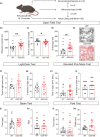
FIGURE 1
FIGURE 1
Assessment of motor and non‐motor behaviors in WT and G2019S mice. Flow‐chart illustrating the experimental design (a). Spontaneous horizontal locomotion and rearing were measured in the open field test (b–e). Panel (e) shows typical tracks for a WT mouse and a G2019S mouse during the first 10 min of the trial. *p < 0.05; ** p < 0.01 Student's unpaired t‐test; ## p < 0.01 Mann–Whitney test. Anxiety‐like behavior was assessed with the light/dark test (f, g) and the EPM test (h, i). Fine motor coordination and balance were assessed with the beam walking test (j, k) and the pole test (l, m). Results are mean ± s.e.m. from n = 12–20 mice: open field test, WT n = 15, G2019S n = 20; light/dark test, WT n = 12, G2019S n = 14; EPM, WT n = 13, G2019S n = 19; Pole test, WT n = 17, G2019S n = 15; Beam test, WT n = 19, G2019S n = 15
FIGURE 2
FIGURE 2
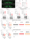
Integrity of SNc‐ and VTA‐DA neurons. (a) Confocal images showing immunofluorescence for TH in the VTA and SNc of a WT mouse and a G2019S mouse (Scale bars 500 μm, 18 z‐stacks with 2 μm interval). (b, c) Number of TH‐positive cells in the SNc and VTA of WT mice (n = 4 mice) and G2019S mice (n = 4 mice). (d–g) Western blotting of TH and DAT in the striatum (n = 24 WT mice and n = 23–27 G2019S mice) and nucleus accumbens (n = 15 WT mice and n = 12–13 G2019S mice). (h–m) Frequency (Hz) and coefficient of variation (CV, %) of the spontaneous firing, measured in cell‐attached mode, of SNc‐ (h, i) and VTA‐DA neurons (k, l) (n = 15–23 cells from four to seven WT mice and n = 23–27 cells from seven G2019S mice). Traces in (j) and (m) are example recordings from SNc‐ and VTA‐DA neurons in WT and G2019S mice
FIGURE 3
FIGURE 3
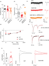
Glutamatergic synaptic transmission in SNc‐DA neurons. Amplitude (a) and frequency (b) of sEPSCs measured in SNc‐DA neurons (n = 10 cells from 4 WT mice, n = 15 cells from 7 G2019S mice in aCSF, and n = 11 cells from 4 G2019S mice in LRRK2‐IN‐1, 1 μM). * p < 0.05, ** p < 0.01 nested one‐way ANOVA followed by multiple comparisons (Tukey). (c) Example recordings from three SNc‐DA neurons in a WT mouse, a G2019S mouse in aCSF, and a G2019S mouse in LRRK2‐IN‐1. Traces on the right are examples of single sEPSCs. (d, e) paired‐pulse ratio of two successive AMPAR‐EPSCs at various interstimulus intervals (n = 10 cells from six WT mice and n = 16 cells from nine G2019S mice). * p < 0.05 nested t‐test. Traces in (e) are examples of recordings of paired EPSCs in WT and G2019S mice evoked at 20 ms and 1000 ms intervals. (f, g) Ratio between AMPAR‐EPSCs and NMDAR‐EPSCs in SNc‐DA neurons (n = 7 cells from 3 WT mice and n = 7 cells from 6 G2019S mice). Traces in (g) are examples of recordings of AMPAR‐EPSCs (recorded at −80 mV) and NMDAR‐EPSCs (recorded at +40 mV)
FIGURE 4
FIGURE 4
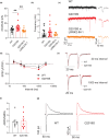
Glutamatergic synaptic transmission in VTA‐DA neurons. Amplitude (a) and frequency (b) of sEPSCs measured in VTA‐DA neurons (n = 20 cells from eight WT mice, n = 19 cells from seven G2019S mice in aCSF, and n = 14 cells from thre G2019S mice in LRRK2‐IN‐1). ** p < 0.01 nested one‐way ANOVA followed by multiple comparisons (Tukey). (c) Example recordings from three VTA‐DA neurons in a WT mouse, a G2019S mouse in aCSF, and a G2019S mouse in LRRK2‐IN‐1. Traces on the right are examples of single sEPSCs. (d, e) paired‐pulse ratio of two successive AMPAR‐EPSCs at various interstimulus intervals (n = 9–13 cells from four WT mice and n = 8–10 cells from eight G2019S mice). Traces in (e) are examples of recordings of paired EPSCs in WT and G2019S mice evoked at 20 ms and 1000 ms intervals. (f, g) Ratio between AMPAR‐EPSCs and NMDAR‐EPSCs in VTA‐DA neurons (n = 8 cells from five WT mice and n = 9 cells from seven G2019S mice). Traces in (g) are examples of recordings of AMPAR‐EPSCs (recorded at −80 mV) and NMDAR‐EPSCs (recorded at +40 mV)
FIGURE 5
FIGURE 5
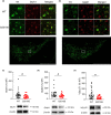
Altered protein amounts of markers of glutamatergic synapses in the ventral midbrain. (a, b) Upper panel: Immunofluorescence detection of TH (green), GluA1 (a, red) and GluN1 (b, red) in the medial SNc of a WT mouse and a G2019S mouse. TH‐positive neurons were co‐labeled with GluA1 (a) and GluN1 (b) (scale bars 50 μm, 18 z‐stacks with 2 μm interval). Lower panel: Lower magnification image indicating, with the white square, the location of the higher magnification upper panels in the medial SNc (Scale bar 500 μm). (c–e) Western blotting of GluA1 (c), GluN1 (d), and VGLUT1 (e) in the ventral midbrain of WT mice (n = 24–25) and G2019S mice (n = 22). # p < 0.05 Mann–Whitney test

References

    1. Beccano‐Kelly, D. A. , Kuhlmann, N. , Tatarnikov, I. , Volta, M. , Munsie, L. N. , Chou, P. , Cao, L. P. , Han, H. , Tapia, L. , Farrer, M. J. , & Milnerwood, A. J. (2014). Synaptic function is modulated by LRRK2 and glutamate release is increased in cortical neurons of G2019S LRRK2 knock‐in mice. Frontiers in Cellular Neuroscience, 8, 301. - PMC - PubMed
    1. Belluzzi, E. , Gonnelli, A. , Cirnaru, M. D. , Marte, A. , Plotegher, N. , Russo, I. , Civiero, L. , Cogo, S. , Carrion, M. P. , Franchin, C. , Arrigoni, G. , Beltramini, M. , Bubacco, L. , Onofri, F. , Piccoli, G. , & Greggio, E. (2016). LRRK2 phosphorylates pre‐synaptic N‐ethylmaleimide sensitive fusion (NSF) protein enhancing its ATPase activity and SNARE complex disassembling rate. Molecular Neurodegeneration, 11, 1. - PMC - PubMed
    1. Belluzzi, E. , Greggio, E. , & Piccoli, G. (2012). Presynaptic dysfunction in Parkinson's disease: A focus on LRRK2. Biochemical Society Transactions, 40, 1111–1116. - PubMed
    1. Brichta, L. , & Greengard, P. (2014). Molecular determinants of selective dopaminergic vulnerability in Parkinson's disease: An update. Frontiers in Neuroanatomy, 8, 152. - PMC - PubMed
    1. Chen, C. , Soto, G. , Dumrongprechachan, V. , Bannon, N. , Kang, S. , Kozorovitskiy, Y. , & Parisiadou, L. (2020). Pathway‐specific dysregulation of striatal excitatory synapses by LRRK2 mutations. eLife, 9, 1–26. - PMC - PubMed

MeSH terms

Substances