Review
. 2022 Jan 28:12:745835.
doi: 10.3389/fmicb.2021.745835. eCollection 2021.

Micro-Technologies for Assessing Microbial Dynamics in Controlled Environments

Affiliations
Review

Micro-Technologies for Assessing Microbial Dynamics in Controlled Environments

Shanna-Leigh Davidson et al. Front Microbiol. .

Abstract

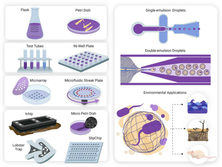
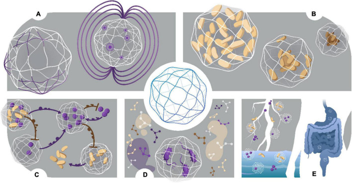
With recent advances in microfabrication technologies, the miniaturization of traditional culturing techniques has provided ideal methods for interrogating microbial communities in a confined and finely controlled environment. Micro-technologies offer high-throughput screening and analysis, reduced experimental time and resources, and have low footprint. More importantly, they provide access to culturing microbes in situ in their natural environments and similarly, offer optical access to real-time dynamics under a microscope. Utilizing micro-technologies for the discovery, isolation and cultivation of "unculturable" species will propel many fields forward; drug discovery, point-of-care diagnostics, and fundamental studies in microbial community behaviors rely on the exploration of novel metabolic pathways. However, micro-technologies are still largely proof-of-concept, and scalability and commercialization of micro-technologies will require increased accessibility to expensive equipment and resources, as well as simpler designs for usability. Here, we discuss three different miniaturized culturing practices; including microarrays, micromachined devices, and microfluidics; advancements to the field, and perceived challenges.

Keywords: microarrays; microfluidics; micromachined devices; nanocultures; unculturable microbes.


Conflict of interest statement

The authors declare that the research was conducted in the absence of any commercial or financial relationships that could be construed as a potential conflict of interest.

Figures

FIGURE 1
FIGURE 1
The miniaturization of cell culturing techniques has revolutionized single-cell studies for culturing microorganisms deemed “unculturable.” Conventional culturing methods (mL—L). Anaerobic jars, flasks and Petri dishes have been used since they were first developed in the 1800s and still form the basis for traditional cell culturing today. Serial dilutions and microwell plates (μL—mL). These mark the first scaling down of culturing techniques and allow for quantification of cell growth and automated screening. Microarray technologies (nL—μL). Microarray printing of droplets allows for highly automated single-cell analyses and investigation of metabolic enzymatic reactions. Two examples include the Microbe Observation and Cultivation Array (MOCA) (Gao et al., 2013) and the Microfluidic Streak Plate (MSP) (Jiang et al., 2016). Micromachined devices (nL—μL). Micromachined devices make use of microfabrication techniques to design devices containing microwells or chambers for cultivating and screening microbes. Examples of such devices include the ichip (Nichols et al., 2010) micro-Petri dish (Ingham et al., 2007), lobster traps (Connell et al., 2010), and SlipChip (Ma et al., 2014). Microfluidic technologies (pL—nL). Microfluidics offer observations with flow dynamics in microfluidic channels, and isolation and compartmentalization of cells in the form of single (Chang et al., 2015) and double (Niepa et al., 2016) emulsion droplets. Droplets can be aqueous or gel phase (Lin et al., 2011), offering tunable constraints for study design. Culturing the “unculturable”- from macroscale to nanoscale. Untapped potential lies awaiting exploration in microbiomes such as marine, animal and soil systems. Discoveries of novel species may influence drug discovery, human health and other fields including bioremediation and agriculture.
FIGURE 2
FIGURE 2
Microarray technology. (A) Microarrays may be stamped or written onto Petri dishes with a microfluidic pen. The hydrophilic droplets containing the cell inoculum are immersed under mineral oil to prevent evaporation and droplet coalescence. Dilution-to-extinction results in compartmentalization of the cells, such that there is one cell per droplet. Competition of resources is negated, allowing slow growers to not be outcompeted. The array is highly amenable to automated screening under a microscope. (B–E) Microfluidic Streak Plate demonstrating isolation and cultivation of bacterial cells from a mixed consortium of RFP- and GFP-tagged E. coli. Cells can be imaged in real-time (D) to show growth dynamics within a single droplet and relative growth (E) of each species can be observed by quantifying fluorescence intensity of each species. Reproduced with permission (Jiang et al., 2016). Copyright 2016, American Society for Microbiology.
FIGURE 3
FIGURE 3
Drug discovery pipeline through culturing the “unculturable.” The ichip was used to culture novel species in an environmental soil sample (Ling et al., 2015). Species that successfully passaged onto agar plates were used to screen for antibiotic activity against S. aureus. Bioactive compounds were extracted, purified, characterized, and tested in vitro against a multitude of pathogenic organisms. This pipeline led to the discovery of teixobactin, a novel antibiotic compound that showed no mutants acquiring resistance against it in preliminary testing. After discovery, novel drugs go through rigorous safety and efficacy testing before they may be developed into safe consumer products. Novel discoveries such as this show the untapped potential of “Microbial dark matter”.
FIGURE 4
FIGURE 4
Nanocultures provide an ideal environment to study microbial growth dynamics over spatial-temporal scales that recapitulate that of macroscale flask cultures. Growth of P. aeruginosa was observed over the course of 20 h, whereby exponential growth is achieved between 7 and 13 h and stationary phase is attained after 13 h of incubation. The nanocultures shrink in size due to consumption of resources, and the volumetric flowrate of water leaving the capsules may be calculated at each stage by observing the decrease in capsule diameter in real-time (Usman et al., 2021).
FIGURE 5
FIGURE 5
Characterization and applications of nanocultures. (A) Chemical and Magnetic functionalization. Nanocultures are custom designed to fit specific applications. Addition of functional group DMAA into the polymer membrane increases free volume, changing the selective permeability properties of the membrane. Similarly, addition of magnetic oxide allows for easy retrieval after nanocultures are freely suspended in an environmental sample. To collect nanocultures, a magnet is simply moved over the sample. (B) Investigation of osmotic stress. Nanocultures may be used to study single cell and/or community response to physical insults, such as osmotic stress. (C) Biochemical Interactions. Intra- and Inter-species dynamics may be studied in real time, whereby the semi-permeable membrane provides physical containment of the cells but allows for cross signaling between nanocultures in the form of small molecules. Furthermore, secreted small molecules may be studied for biological relevance, such as drug discovery or beneficial secondary metabolites for symbiotic relationships. (D) Growth of fastidious species. Growth of fastidious species has been demonstrated by culturing C. difficile under a microscope, negating the use of anaerobic jars or chambers. (E) Culturing the “unculturable.” Nanocultures can be used to successfully culture the “unculturable” from many environmental sources, such as soil, marine and human microbiomes.

References

    1. Aleklett K., Kiers E. T., Ohlsson P., Shimizu T. S., Caldas V. E. A., Hammer E. C. (2018). Build your own soil: exploring microfluidics to create microbial habitat structures. ISME J. 12 312–319. 10.1038/ismej.2017.184 - DOI - PMC - PubMed
    1. Alkayyali T., Pope E., Wheatley S. K., Cartmell C., Haltli B., Kerr R. G., et al. (2021). Development of a microbe domestication pod (MD Pod) for in situ cultivation of micro-encapsulated marine bacteria. Biotechnol. Bioeng. 118 1166–1176. 10.1002/bit.27633 - DOI - PubMed
    1. Andersson H., van den Berg A. (2004). Microtechnologies and nanotechnologies for single-cell analysis. Curr. Opin. Biotechnol. 15 44–49. 10.1016/j.copbio.2004.01.004 - DOI - PubMed
    1. Banks P. (2009). The Microplate Market Past, Present and Future [Online]. Drug Discovery World. Available online at: https://www.ddw-online.com/the-microplate-market-past-present-and-future... (accessed 01 04, 2021)
    1. Barkal L. J., Theberge A. B., Guo C.-J., Spraker J., Rappert L., Berthier J., et al. (2016). Microbial metabolomics in open microscale platforms. Nat. Commun. 7:10610. 10.1038/ncomms10610 - DOI - PMC - PubMed

LinkOut - more resources