Case Reports
. 2022 Feb 3:2022:8076649.
doi: 10.1155/2022/8076649. eCollection 2022.

Syringocystadenocarcinoma Papilliferum in a Fifteen-Year-Old Girl: A Case Report and Review of the Literature

Affiliations
Case Reports

Syringocystadenocarcinoma Papilliferum in a Fifteen-Year-Old Girl: A Case Report and Review of the Literature

Jordan N Halsey et al. Case Rep Dermatol Med. .

Abstract

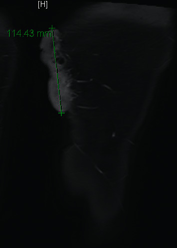
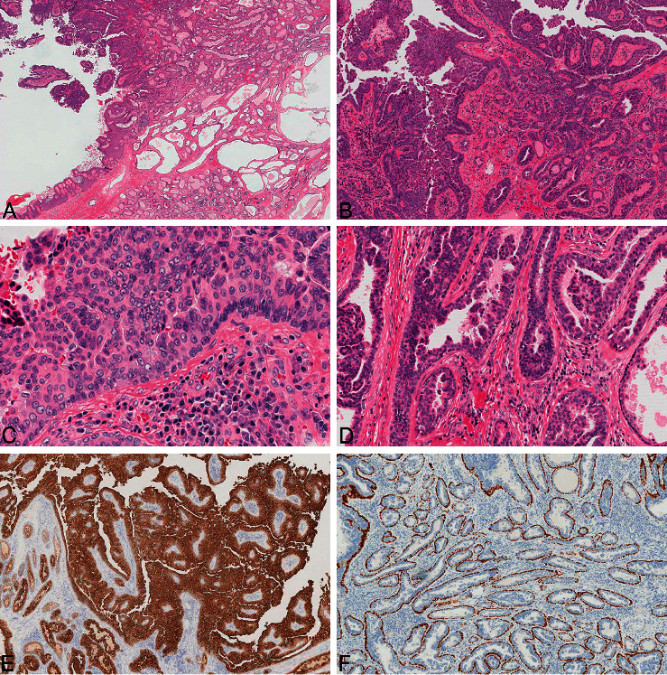
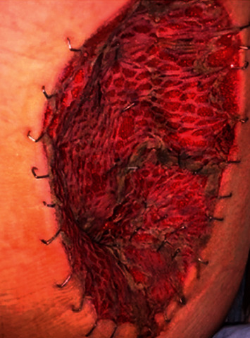
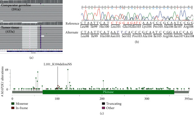
Syringocystadenocarcinoma papilliferum (SCACP) is a rare malignant neoplasm arising from adnexal tissues and is the malignant complement to the benign neoplasm syringocystadenoma papilliferum (SCAP). SCACP lesions appear as raised nodules or inflammatory plaques and can be associated with SCAP or nevus sebaceous. There have been fewer than 100 described cases of this neoplasm in the literature, and all previously published cases have been described in adults, with the majority occurring in the elderly. We present a case of an adolescent female with a syringocystadenocarcinoma papilliferum arising from a large thigh mass harboring an in-frame alteration in MAP2K1 along with a brief review of the literature.


Conflict of interest statement

The authors declare no conflicts of interest.

Figures

Figure 1
Figure 1
Left posterior thigh pedunculated mass.
Figure 2
Figure 2
MRI imaging of the left thigh showing posteromedial mass, with arterial blood supply from branches of the deep femoral artery.
Figure 3
Figure 3
The tumor was composed of both exophytic and underlying infiltrative components, the former showing a papillary pattern similar to syringocystadenoma papilliferum and the latter showing closely spaced tubular glands similar to tubular apocrine adenoma (a). Florid overgrowth of the outer epithelial cells produced complex, maze-like spaces between epithelial cells and cellular tufts off the surface of the tumor (b). The outer epithelial cells showed crowded, overlapping, atypical nuclei with prominent nucleoli with mitotic activity (c). The inner luminal cells of the dermal glands demonstrated bulbous, micropapillary projections (d). CK7 was diffusely expressed by both outer epithelial cells of the papillae and inner luminal cells of the dermal glands (e). P63-positive basal cells bordered the papillae and glands (f).
Figure 4
Figure 4
Postoperative image following reresection with margins and skin graft placement.
Figure 5
Figure 5
(a) Integrative genomics viewer image of aligned sequencing reads for exon 3 of MAP2K1 derived from enhanced exome sequencing. The top panel shows the comparator germline (peripheral blood) with the absence of the complex indel at an average of 591x depth for this region. The bottom panel shows the tumor tissue demonstrating an in-frame complex indel with an average of 433x depth for this region. (b) Sanger sequencing chromatogram of the in-frame MAP2K1 (NM_002755.4) complex indel (c.301_311delinsAATTC:p.Leu101_Lys104delinsAsnSer). The corresponding amino acid sequence is described below the genomic sequence. Italicized reference sequence in red indicates the deletion, and the italicized alternate sequence in blue indicates the insertion. (c) Lollipop plot of MAP2K1 variants deposited in cBioPortal. The p.Leu101_Lys104delinsAsnSer described in this report has not been previously reported.

References

    1. Lee K. G., Choi W., Lim J. S., Hahn H. J., Myung K. B., Cheong S. H. Syringocystadenocarcinoma papilliferum: a case report and review of the literature. Annals of Dermatology . 2019;31(5):559–562. doi: 10.5021/ad.2019.31.5.559. - DOI - PMC - PubMed
    1. Bondi R., Urso C. Syringocystadenocarcinoma papilliferum. Histopathology . 1996;28(5):475–477. doi: 10.1046/j.1365-2559.1996.t01-4-297345.x. - DOI - PubMed
    1. Castillo L., Moreno A., Tardío J. C. Syringocystadenocarcinoma papilliferum in situ: report of a case with late recurrence. The American Journal of Dermatopathology . 2014;36(4):348–352. doi: 10.1097/dad.0b013e3182a38bb9. - DOI - PubMed
    1. Kazakov D. V., Requena L., Kutzner H., et al. Morphologic diversity of syringocystadenocarcinoma papilliferum based on a clinicopathologic study of 6 cases and review of the literature. The American Journal of Dermatopathology . 2010;32(4):340–347. doi: 10.1097/dad.0b013e3181b96c0c. - DOI - PubMed
    1. Zhang Y., Kong Y.-Y., Cai X., Shen X.-X., Kong J.-C. Syringocystadenocarcinoma papilliferum: clinicopathologic analysis of 10 cases. Journal of Cutaneous Pathology . 2017;44(6):538–543. doi: 10.1111/cup.12934. - DOI - PubMed

Publication types

LinkOut - more resources