. 2022 Feb 7;11(1):2034355.
doi: 10.1080/2162402X.2022.2034355. eCollection 2022.

Trispecific T-cell engagers for dual tumor-targeting of colorectal cancer

Affiliations

Trispecific T-cell engagers for dual tumor-targeting of colorectal cancer

Antonio Tapia-Galisteo et al. Oncoimmunology. .

Abstract

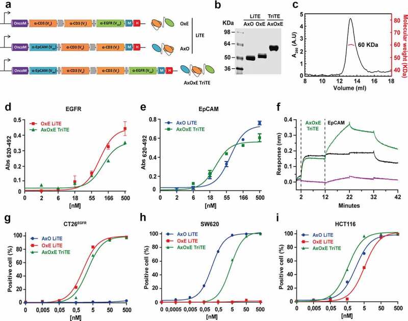
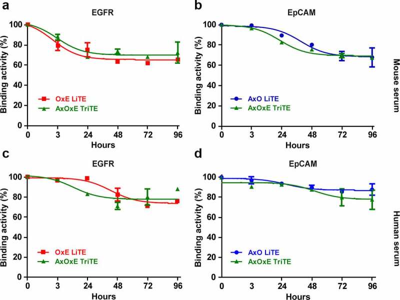
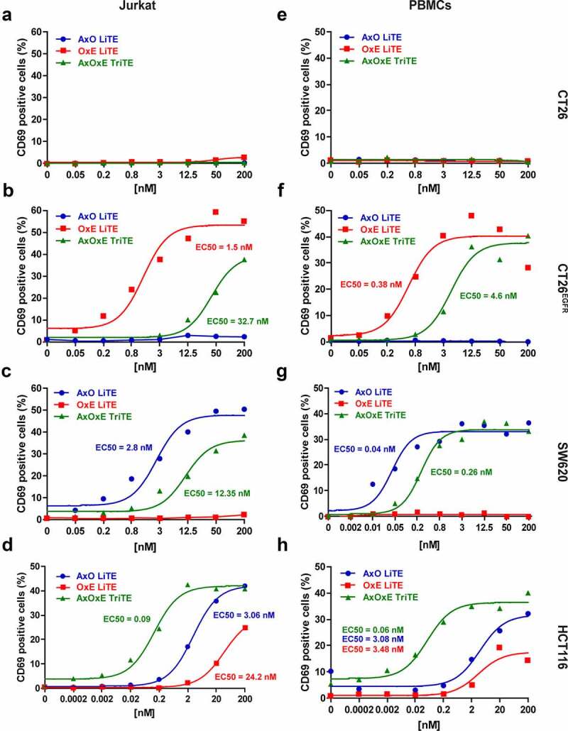
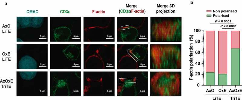
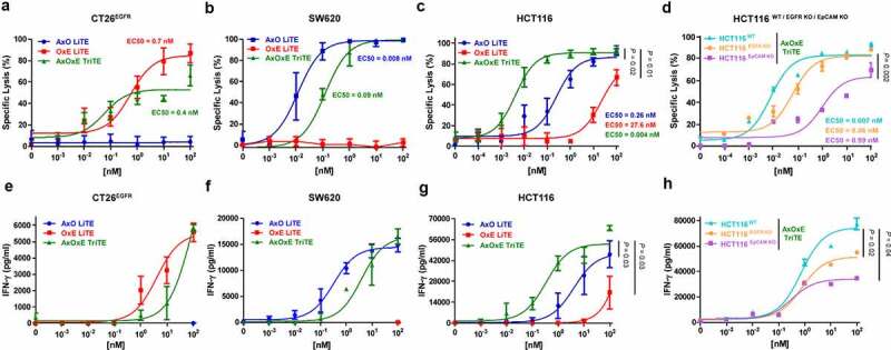
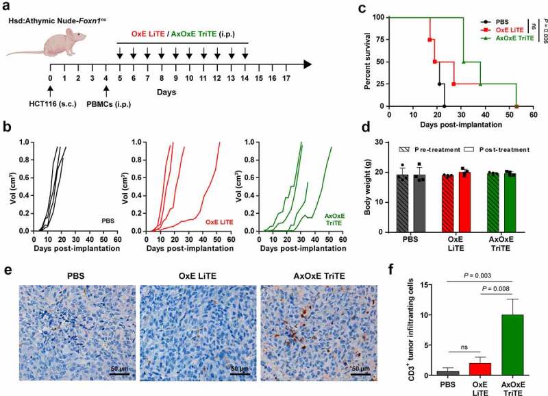
Retargeting of T lymphocytes toward cancer cells by bispecific antibodies has demonstrated its therapeutic potential, with one such antibody approved for the treatment of acute lymphoblastic leukemia (blinatumomab) and several other in clinical trials. However, improvement of their efficacy and selectivity for solid tumors is still required. Here, we describe a novel tandem T-cell recruiting trispecific antibody for the treatment of colorectal cancer (CRC). This construct, termed trispecific T-cell engager (TriTE), consists of a CD3-specific single-chain Fv (scFv) flanked by anti-epidermal growth factor receptor (EGFR) and anti-epithelial cell adhesion molecule (EpCAM) single-domain VHH antibodies. The TriTE was well expressed in mammalian and yeast cells, bound the cognate antigens of the three parental antibodies, and enabled the specific cytolysis of EGFR- and/or EpCAM-expressing cancer cells, without inducing T cell activation and cytoxicity against double-negative (EGFR-EpCAM-) cancer cells. Bivalent bispecific targeting of double-positive HCT116 cells by TriTE improved in vitro potency up to 100-fold compared to single-positive cells and significantly prolonged survival in vivo. In addition, it was less efficient at killing single-positive target cells than the corresponding bispecific controls, leading to potentially enhanced tumor specificity. Moreover, dual targeting of two tumor-associated antigens may contribute toward preventing the tumor escape by antigen loss caused by selective pressures from conventional single-targeting T-cell engagers, and may help to overcome antigenic heterogeneity.

Keywords: Trispecific antibodies; cancer immunotherapy; colorectal cancer; scFv; single-domain antibodies; tandem antibodies.


Conflict of interest statement

No potential conflicts of interest were disclosed.

Figures

Figure 1.
Figure 1.
Schematic representation and characterization of trispecific T-cell engager (TriTE) and corresponding light T-cell engagers (LiTEs). (a) Genetic structure of the tandem VHH-scFv AxO LiTE formed by fusing the anti-EpCAM A2 VHH (blue box) N-terminally to the CD3-specific OKT3 scFv (Orange box); the scFv-VHH OxE LiTE comprising the anti-EGFR EGa1 VHH (green box) fused C-terminally to the OKT3 scFv; and the VHH-scFv-VHH AxOxE TriTE with anti-EpCAM and anti-EGFR VHH fused to the N- and C-terminus of OKT3 scFv, respectively. The Oncostatin M signal peptide (purple box) is used to direct secretion of recombinant antibody, and the myc/6xHis tags (dark blue and red boxes) were appended for immunodetection and affinity purification, respectively. Schematic representations showing arrangement of VHH and scFv in each construct are shown on the right. (b) Reducing SDS-PAGE of the three constructs and (c) SEC analysis of the purified AxOxE TriTE with the indicated molecular weight measured at the center of the chromatography peak (red line). (d-e) Titration ELISA against plastic-immobilized EGFR and EpCAM. Experiments were performed at least twice in duplicates. Mean ± SD are shown at each concentration. (f) Biolayer interferometry (BLI)-derived sensorgrams for the interaction between immobilized EGFR and AxOxE TriTE in the presence (green) or not (black) of soluble EpCAM. Note that the TriTE was present during association with EpCAM to prevent dissociation of the TriTE from immobilized EGFR. (g-i) FACS on CT26EGFR (EGFR+EpCAM), SW620 (EGFREpCAM+) and HCT116 cells (EGFR+EpCAM+). Percentages of positive cells are shown at each concentration. AxO = A2 (anti-EpCAM VHH) x OKT3 (anti-CD3 scFv); OxE = OKT3 x Ega1 (anti-EGFR VHH); AxOxE = A2 x OKT3 x Ega1.
Figure 2.
Figure 2.
Serum stability of purified EpCAMxCD3xEGFR TriTE and EpCAMxCD3 and CD3xEGFR LiTEs. AxOxE TriTE and LiTEs were incubated in mouse (a-b) or human (c-d) serum at 37°C for 96 hours and their functional activity was analyzed by ELISA against plastic-immobilized EGFR (left) or EpCAM (right). Experiments were performed twice in duplicates. Mean ± SD are shown at each time point. AxO = A2 (anti-EpCAM VHH) x OKT3 (anti-CD3 scFv); OxE = OKT3 x Ega1 (anti-EGFR VHH); AxOxE = A2 x OKT3 x Ega1.
Figure 3.
Figure 3.
Induction of T cell activation by purified EpCAMxCD3xEGFR TriTE and EpCAMxCD3 and CD3xEGFR LiTEs. (a,e) CT26 cells (EGFREpCAM); (b,f) CT26EGFR cells (EGFR+EpCAM); (c,g) SW620 cells (EGFREpCAM+) or (d,h) HCT116 cells (EGFR+EpCAM+) were cocultured with Jurkat cells (left) or PBMCs (right) at the effector/target (E/T) ratio of 5:1 in the presence of different concentrations of purified AxOxE TriTE and LiTEs. After 24 hours, the surface expression of T cell activation marker CD69 was determined by FACS analysis. EC50 values are provided according to the color code. Experiments were performed three times, one representative experiment is shown. AxO = A2 (anti-EpCAM VHH) x OKT3 (anti-CD3 scFv); OxE = OKT3 x Ega1 (anti-EGFR VHH); AxOxE = A2 x OKT3 x Ega1.
Figure 4.
Figure 4.
Immunological synapse formation is triggered by EpCAMxCD3xEGFR TriTE. (a) Images of immunological synapse (IS) assembly by Jurkat cells co-cultured with EpCAM+EGFR+ HCT116 cells (cyan) in the presence of AxOxE TriTE or LiTEs. The green (CD3ε) and red (F-actin) channels, as well as the merged images, are shown. The IS topology obtained from the 3D reconstructions of regions of interest in confocal stacks (white square) containing the red and the green channels is shown on the right. Experiments were performed three times; results of one representative experiment are shown. Scale bar 5 µm. (b) Percentages of T cells showing F-actin polarization at the IS in each condition are shown. Statistical differences were examined by two-coiled chi-square test. AxO = A2 (anti-EpCAM VHH) x OKT3 (anti-CD3 scFv); OxE = OKT3 x Ega1 (anti-EGFR VHH); AxOxE = A2 x OKT3 x Ega1.
Figure 5.
Figure 5.
Specific cytotoxicity and IFN-γ secretion elicited by purified EpCAMxCD3xEGFR TriTE and EpCAMxCD3 and CD3xEGFR LiTEs. CT26EGFR-Luc cells (a,e), SW620Luc cells (b,f) or HCT116Luc (c,g) cells were cocultured in 96-well plates with PBMCs at the effector/target (E/T) ratio of 5:1 in the presence of different concentrations of purified AxOxE TriTE and LiTEs. In additional experiments, HCT116Luc were compared with the corresponding EGFR and EpCAM knockout cell lines in the presence of TriTE serial dilutions (d,h). After 72 hours, specific cytolysis of tumor cells were measured by bioluminescence assay (upper) and IFN-γ production was determined in CM by ELISA (lower). Percent specific lysis was calculated relative to an equal number of tumor cells cultured with PBMCs in the absence of purified antibodies. EC50 values are provided according to the color code. PBMCs were obtained from 3 different donors, and experiments were performed in triplicate. Statistical differences were examined by unpaired Student’s t-test assuming a normal distribution. Results are expressed as a mean ± SD. AxO = A2 (anti-EpCAM VHH) x OKT3 (anti-CD3 scFv); OxE = OKT3 x Ega1 (anti-EGFR VHH); AxOxE = A2 x OKT3 x Ega1.
Figure 6.
Figure 6.
In vivo therapeutic effect of EpCAMxCD3xEGFR TriTE and CD3xEGFR LiTE. (a) Hsd:Athymic Nude-Foxn1nu mice were inoculated subcutaneously with 2 × 106 HCT116 tumor cells. Mice were randomized into groups (n = 4/group) when tumors reached 0.2 cm in diameter and injected intraperitoneally with 107 freshly isolated PBMCs. Then, mice were treated with intraperitoneally injections of PBS, OxE LiTE or AxOxE TriTE daily. (b) Tumor volume growth curves for individual mice are represented. (c) Kaplan-Meier survival curves of AxOxE TriTE- and OxE LiTE-treated mice, log-rank (Mantel-Cox) test. (d) Mice weight before and after treatment are shown. (e) Representative images of CD3+ TIL immunostaining in tissue sections from HCT116 tumors treated with PBS, OxE LiTE or AxOxE TriTE. Tumors were resected at termination of the experiment shown above. Error bar = 50 µm. (f) Quantification of CD3+ TILs in three independent fields/tissue section. Statistical differences were examined by unpaired Student’s t-test assuming a normal distribution. Results are expressed as a mean ± SD. OxE = OKT3 (anti-CD3 scFv) x Ega1 (anti-EGFR VHH), AxOxE = A2 (anti-EpCAM VHH) x OKT3 x Ega1.

References

    1. Kontermann RE, Brinkmann U.. Bispecific Antibodies. Drug Discov Today. 2015;20(7):838–14. doi:10.1016/j.drudis.2015.02.008. - DOI - PubMed
    1. Przepiorka D, Ko C-W, Deisseroth A, Yancey CL, Candau-Chacon R, Chiu H-J, Gehrke BJ, Gomez-Broughton C, Kane RC, Kirshner S, et al. FDA Approval: blinatumomab. Clin Cancer Res. 2015;21(18):4035–4039. doi:10.1158/1078-0432.CCR-15-0612. - DOI - PubMed
    1. Suurs FV, Lub-de Hooge MN, de Vries EGE, de Groot DJA. A Review of Bispecific Antibodies and Antibody Constructs in Oncology and Clinical Challenges. Pharmacol Ther. 2019;201:103–119. doi:10.1016/j.pharmthera.2019.04.006. - DOI - PubMed
    1. Compte M, Harwood SL, Muñoz IG, Navarro R, Zonca M, Perez-Chacon G, Erce-Llamazares A, Merino N, Tapia-Galisteo A, Cuesta AM, et al. A Tumor-Targeted Trimeric 4-1BB-Agonistic Antibody Induces Potent Anti-Tumor Immunity without Systemic Toxicity. Nat Commun. 2018;9(1):4809. doi:10.1038/s41467-018-07195-w. - DOI - PMC - PubMed
    1. Gauthier L, Morel A, Anceriz N, Rossi B, Blanchard-Alvarez A, Grondin G, Trichard S, Cesari C, Sapet M, Bosco F, et al. Multifunctional Natural Killer Cell Engagers Targeting NKp46 Trigger Protective Tumor Immunity. Cell. 2019;177(7):1701–1713.e16. doi:10.1016/j.cell.2019.04.041. - DOI - PubMed

Publication types

Substances