

Role of Magnetic Resonance Imaging in the Evaluation of Rotator Cuff Tears
- PMID: 35154995
- PMCID: PMC8819335
- DOI: 10.7759/cureus.21025
Role of Magnetic Resonance Imaging in the Evaluation of Rotator Cuff Tears
Abstract
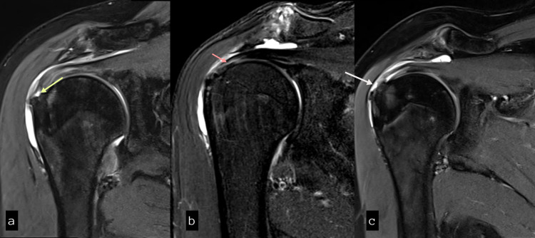
Background Magnetic resonance imaging (MRI), with the advent of surface coils, is becoming the modality of choice for imaging soft tissues around the shoulder joint. Good knowledge regarding the MR characteristics of rotator cuff tendons, acromion, and the abnormalities in these tendons is necessary for appropriate diagnosis. Methods This was a hospital-based descriptive, analytical and prospective study conducted at our tertiary care hospital. The study was performed on 50 patients with rotator cuff lesions detected on MRI of the shoulder joint. Results The age distribution found in the study is between 19 and 66 years with mean being 43 ± 14.8 years. The peak incidence was found in the fifth and sixth decades of life. Gender-wise distribution of rotator cuff pathologies has shown no significant gender variation. The pain was the most common presenting complaint. An abnormal supraspinatus tendon was seen in 82% of the 50 study patients, making it the most commonly affected tendons, followed by subscapularis and infraspinatus tendons. No apparent teres minor pathology was identified in the study patients. The most common pathology affecting the supraspinatus tendon was tendinosis (38%) closely followed by a partial tear (36%). Among the partial tears, the articular surface type of tear was the most common. About 52% patients had type II (curved) acromion; making it the most common type of acromion followed by type III (hook), supraspinatus tendinopathy was more common in type II acromion. A reduction in the acromiohumeral distance can cause supraspinatus tendinosis and also makes it more susceptible to tear. About 45.5% showed supraspinatus tendon tears when the acromiohumeral distance was less than 8mm as compared to 13.6% when more than 10mm. Only 4.2% had normal supraspinatus tendon in patients with this distance less than 7mm. Conclusion MRI provides valuable information to the orthopaedic surgeon regarding the status of tendons, bones, and joints. In order to choose the appropriate course of action, it is crucial first to identify the issue and report relevant data from rotator cuff imaging. A full grasp of the rotator cuff's architecture and function, as well as the repercussions of rotator cuff diseases, is required.
Keywords: acromiohumeral distance; acromion; rotator cuff tears; shoulder mri; supraspinatus.
Copyright © 2022, Koganti et al.
Conflict of interest statement
The authors have declared that no competing interests exist.
Figures


References
-
- Wong M, Kiel J. Europe PMC. Treasure Island, FL: StatPearls Publishing; 2021. Anatomy, Shoulder and Upper Limb, Acromioclavicular Joint. - PubMed
-
- Rotator cuff: evaluation with US and MR imaging. Seibold CJ, Mallisee TA, Erickson SJ, Boynton MD, Raasch WG, Timins ME. Radiographics. 1999;19:685–705. - PubMed
-
- MR imaging of rotator cuff injury: what the clinician needs to know. Morag Y, Jacobson JA, Miller B, De Maeseneer M, Girish G, Jamadar D. Radiographics. 2006;26:1045–1065. - PubMed
-
- Accuracy of MRI, MR arthrography, and ultrasound in the diagnosis of rotator cuff tears: a meta-analysis. de Jesus JO, Parker L, Frangos AJ, Nazarian LN. AJR Am J Roentgenol. 2009;192:1701–1707. - PubMed
-
- High resolution surface coil magnetic resonance imaging of the joints: anatomic correlation. Middleton WD, Macrander S, Lawson TL, et al. Radiographics. 1987;7:645–683. - PubMed
LinkOut - more resources
Full Text Sources