Review
. 2020 Sep 10;83(1):1-10.
doi: 10.1055/s-0040-1716688. eCollection 2022 Feb.

Beyond Antoni: A Surgeon's Guide to the Vestibular Schwannoma Microenvironment

Affiliations
Review

Beyond Antoni: A Surgeon's Guide to the Vestibular Schwannoma Microenvironment

Cathal J Hannan et al. J Neurol Surg B Skull Base. .

Abstract

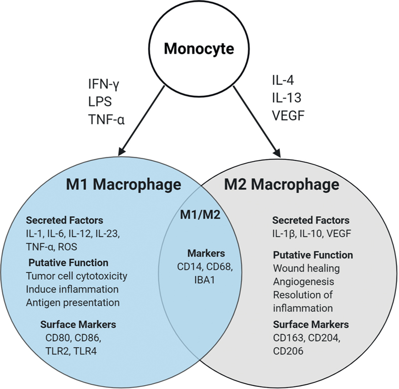
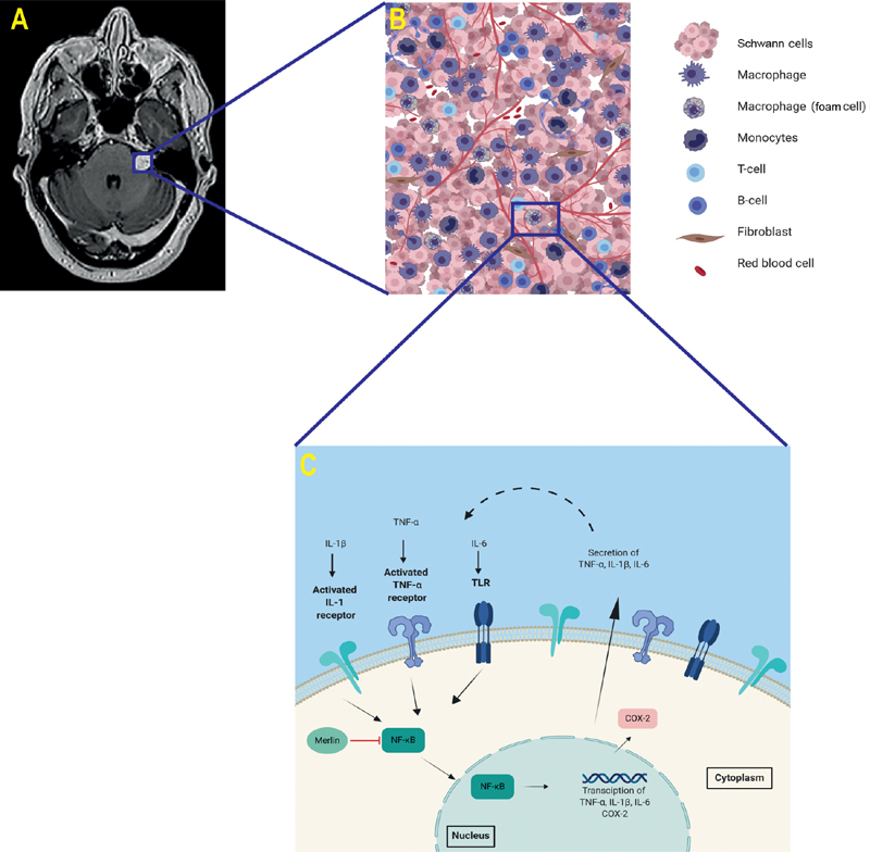
Introduction Vestibular schwannomas (VS) are histologically benign tumors arising from cranial nerve VIII. Far from a homogenous proliferation of Schwann cells, mounting evidence has highlighted the complex nature of the inflammatory microenvironment in these tumors. Methods A review of the literature pertaining to inflammation, inflammatory molecular pathways, and immune-related therapeutic targets in VS was performed. Relevant studies published up to June 2020 were identified based on a literature search in the PubMed and MEDLINE databases and the findings were synthesized into a concise narrative review of the topic. Results The VS microenvironment is characterized by a dense infiltrate of inflammatory cells, particularly macrophages. Significantly higher levels of immune cell infiltration are observed in growing versus static tumors, and there is a demonstrable interplay between inflammation and angiogenesis in growing VS. While further mechanistic studies are required to ascertain the exact role of inflammation in angiogenesis, tumor growth, and Schwann cell control, we are beginning to understand the key molecular pathways driving this inflammatory microenvironment, and how these processes can be monitored and targeted in vivo . Conclusion Observational research has revealed a complex and heterogeneous tumor microenvironment in VS. The functional landscape and roles of macrophages and other immune cells in the VS inflammatory infiltrate are, however, yet to be established. The antiangiogenic drug bevacizumab has shown the efficacy of targeted molecular therapies in VS and there is hope that agents targeting another major component of the VS microenvironment, inflammation, will also find a place in their future management.

Keywords: angiogenesis; antiangiogenic; bevacizumab; biomarkers; immunomodulation; immunotherapy; inflammation; tumor immunology; tumor-associated macrophages; vestibular schwannoma.


Conflict of interest statement

Conflict of Interest D.G.E. reports personal fees from Astrazeneca, outside the submitted work. All the other authors report no conflict of interest.

Figures

Fig. 1
Fig. 1
Macrophage polarity. Typically, monocytes/macrophages can be polarized to an M1 phenotype by Toll like receptor (TLR) ligands such as lipopolysaccharide (LPS), interferon-g (IFN-g), or tumor necrosis factor-a (TNF-a), and secrete proinflammatory cytokines to be antitumorigenic. Alternatively, they can be polarized to an M2 phenotype by vascular endothelial growth factor (VEGF), Interleukin-4 (IL-4) and IL-13, which secrete anti-inflammatory cytokines to be pro-tumorigenic. CD14 and CD68 act as cell surface markers for macrophages, common to M1 and M2 types. Although this dichotomous polarity is somewhat of an oversimplification, the concept of pro and anti-tumoral activity of macrophages is highly clinically relevant. Readers interested in exploring this topic in more detail are directed to an excellent review by Mantovani et al.
Fig. 2
Fig. 2
Overview of biology of vestibular schwannoma (VS). ( A ) An axial T1-W Post-Gd MRI of a VS. ( B ) Magnified image depicting a schematic representation of the tumor microenvironment and the cell populations implicated in the VS microenvironment ( C ) Schematic representation of a positive feedback loop of inflammation in VS. Cytokines bind to cell surface receptors of Schwann's cells, activating the transcription factor NF-κB which demonstrably upregulates TNF-α, IL-1β, IL-6 and COX-2 expression. The cytoskeleton tumor suppressor protein merlin blocks NF-κB activity. COX-2 production is upregulated by NF-κB. Dashed line illustrates the purported positive feedback loop perpetuating the inflammation within VS. COX, cyclooxygenase; Gd, gadolinium; IL, Interleukin; MRI, magnetic resonance imaging; NF, nuclear factor; TNF, tumor necrosis factor; W, weighted.
Fig. 3
Fig. 3
Relationship between hypoxia, angiogenesis and inflammation in VS. ( A ) Immunostains from a growing NF2-related VS demonstrating macroscopic spatial colocalization between Iba1 (ionized calcium-binding adapter molecule 1, marker of active macrophages) and regions of high CD31+ (expressed on endothelial cells, indicative of angiogenesis) microvessel density. Left: Iba1, red, immunoperoxidase, whole mount; CD31, brown, immunoperoxidase, whole mount. ( B ) Higher magnification (×10 HPF) of the area framed in the whole mounts) demonstrating an intratumoral region of high Iba1 + TAM density and high CD31 + microvessel density respectively. Top: Iba1, red; bottom: CD31, brown (immunoperoxidase, ×10). ( C ) Higher magnification (×20 HPF) immunofluoresence images of the area framed in the whole mounts demonstrating areas of cellular colocalization between Iba1 and VEGF (yellow). From left to right: Iba1 (red), VEGF (green) and VEGF-Iba1 (red-green) (immunofluorescence, ×20). HPF, high power field; NF2, neurofibromatosis type 2; VEGF, vascular endothelial growth factor; TAM, tumor-associated macrophages; VS, vestibular schwannoma.
Fig. 4
Fig. 4
Imaging biomarkers of intratumoral inflammation in VS. Representative imaging and histology from a patient with a static VS ( A ); and a growing VS ( B ) is shown. From left to right: axial T1-W post-Gd; parametric map of [ 11 C]-( R )-PK11195 PET specific binding (BP ND ); map of DCE-MRI derived K trans ; and immunostains demonstrating Iba1 (Iba1 red, immunoperoxidase) TAM density in each tumor. Within the growing VS there is demonstrably higher binding of the inflammation PET tracer, [ 11 C]-( R )-PK11195, and elevated K trans suggesting indicating increased vascular density/permeability compared with the static tumor. Comparative immunohistochemistry (Iba1 red, immunoperoxidase) demonstrates that within the growing VS, there is an abundance of intratumoral Iba1 + macrophages. DEC-MRI, dynamic contrast-enhanced magnetic resonance imaging; Gd, gadolinium; PET, positron emission tomography; TAM, tumor-associated macrophages; VS, vestibular schwannoma; W, weighted.

References

    1. Evans D G, Moran A, King A, Saeed S, Gurusinghe N, Ramsden R. Incidence of vestibular schwannoma and neurofibromatosis 2 in the North West of England over a 10-year period: higher incidence than previously thought. Otol Neurotol. 2005;26(01):93–97. - PubMed
    1. Halliday D, Parry A, Evans D G. Neurofibromatosis type 2 and related disorders. Curr Opin Oncol. 2019;31(06):562–567. - PubMed
    1. Pathmanaban O N, Sadler K V, Kamaly-Asl I D. Association of genetic predisposition with solitary schwannoma or meningioma in children and young adults. JAMA Neurol. 2017;74(09):1123–1129. - PMC - PubMed
    1. Lees K A, Tombers N M, Link M J. Natural history of sporadic vestibular schwannoma: a volumetric study of tumor growth. Otolaryngol Head Neck Surg. 2018;159(03):535–542. - PubMed
    1. Stangerup S E, Caye-Thomasen P, Tos M, Thomsen J. The natural history of vestibular schwannoma. Otol Neurotol. 2006;27(04):547–552. - PubMed