

Novel and Specific MRI Features Indicate the Clinical Features of Patients With Rare Hepatic Tumor Epithelioid Hemangioendothelioma
- PMID: 35155220
- PMCID: PMC8828502
- DOI: 10.3389/fonc.2022.729177
Novel and Specific MRI Features Indicate the Clinical Features of Patients With Rare Hepatic Tumor Epithelioid Hemangioendothelioma
Abstract
Objective: To investigate the MRI features and clinical significance of hepatic epithelioid hemangioendothelioma (HEHE).
Methods: Clinical records and MRI findings were retrospectively evaluated in nine HEHE patients from May 2010 to January 2020.
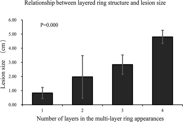
Result: There were 121 lesions in nine patients with a predominantly peripheral distribution. Five lesions (4.13%) in two patients (22.22%) had evidence of capsular retraction, and three patients had lung metastasis (33.33%). Dynamic contrast-enhanced MRI showed progressive enhancement, mainly in two ways: ring enhancement with hypovascularity in four patients (44.44%) and ring enhancement with hypervascularity in five patients (55.56%). Imaging demonstrated a multilayer ring appearance, which was typically observed on T2-weighted imaging (T2WI). The most common appearance consisted of two layers of varying signal, with some images displaying up to four layers. There were significant differences in the size of lesions between different layers of multilayer ring appearance (p < 0.001). All lesions exhibited a two-layer appearance on diffusion-weighted imaging (DWI), with hyperintensity at the periphery and a slightly high signal at the center (except for those with a single layer on T2WI). The "vascular penetration sign" was observed in most lesions, and the blood vessels of 112 lesions (92.56%) were portal vein branches, and five (4.13%) were hepatic vein branches. Pulmonary metastasis was found in three patients with the "vascular penetration sign" of hepatic vein branches.
Conclusion: The multilayer ring appearance on T2WI, the "vascular penetration sign", and the two enhancement patterns may be of great significance in the diagnosis and treatment of HEHE. The "vascular penetration sign" of hepatic vein branches may indicate extrahepatic metastasis.
Keywords: dynamic contrast-enhanced magnetic resonance imaging (MRI); extrahepatic metastasis; hepatic epithelioid hemangioendothelioma (HEHE); hepatic vein branches; liver tumor.
Copyright © 2022 Zhang, Zhang, Zhong, Zhang, Kong, Yu, Chen, Bai, Zhu, Yang and Gao.
Conflict of interest statement
The authors declare that the research was conducted in the absence of any commercial or financial relationships that could be construed as a potential conflict of interest.
Figures


Similar articles
-
[MRI manifestations of 40 cases with the hepatic epithelioid hemangioendothelioma classification based on the morphology and size].Zhonghua Gan Zang Bing Za Zhi. 2024 Jun 20;32(6):545-550. doi: 10.3760/cma.j.cn501113-20230910-00100. Zhonghua Gan Zang Bing Za Zhi. 2024. PMID: 38964897 Chinese.
-
Spectrum of appearances on CT and MRI of hepatic epithelioid hemangioendothelioma.BMC Gastroenterol. 2015 Jun 19;15:69. doi: 10.1186/s12876-015-0299-x. BMC Gastroenterol. 2015. PMID: 26088585 Free PMC article.
-
CT and MRI diagnosis of hepatic epithelioid hemangioendothelioma.Hepatobiliary Pancreat Dis Int. 2010 Apr;9(2):154-8. Hepatobiliary Pancreat Dis Int. 2010. PMID: 20382586
-
Magnetic Resonance Imaging of Uncommon Hepatic Mesenchymal Tumours: Haemangioendothelioma and Angiosarcoma.Curr Med Imaging Rev. 2019;15(4):362-368. doi: 10.2174/1573405614666180628160809. Curr Med Imaging Rev. 2019. PMID: 31989904 Review.
-
Hepatic epithelioid hemangioendothelioma: Update on diagnosis and therapy.World J Clin Cases. 2020 Sep 26;8(18):3978-3987. doi: 10.12998/wjcc.v8.i18.3978. World J Clin Cases. 2020. PMID: 33024754 Free PMC article. Review.
Cited by
-
Specific imaging features indicate the clinical features of patients with hepatic perivascular epithelioid cell tumor by comparative analysis of CT and ultrasound imaging.Front Oncol. 2022 Oct 17;12:908189. doi: 10.3389/fonc.2022.908189. eCollection 2022. Front Oncol. 2022. PMID: 36324566 Free PMC article.
-
Sirolimus combined with interferon-alpha 2b therapy for giant hepatic epithelioid hemangioendothelioma: a case report.Front Oncol. 2022 Aug 24;12:972306. doi: 10.3389/fonc.2022.972306. eCollection 2022. Front Oncol. 2022. PMID: 36081563 Free PMC article.
-
Case report: Successful treatment with the combined therapy of interferon-alpha 2b and anlotinib in a patient with advanced hepatic epithelioid hemangioendothelioma.Front Med (Lausanne). 2022 Dec 2;9:1022017. doi: 10.3389/fmed.2022.1022017. eCollection 2022. Front Med (Lausanne). 2022. PMID: 36530920 Free PMC article.
-
Hepatic Epithelioid Hemangioendothelioma: A Rare Vascular Neoplasm of the Liver.Cureus. 2025 Jul 28;17(7):e88941. doi: 10.7759/cureus.88941. eCollection 2025 Jul. Cureus. 2025. PMID: 40895865 Free PMC article.
-
CT appearances and classification of hepatic epithelioid hemangioendothelioma.Insights Imaging. 2023 Apr 1;14(1):56. doi: 10.1186/s13244-023-01410-z. Insights Imaging. 2023. PMID: 37005950 Free PMC article.
References
LinkOut - more resources
Full Text Sources