

Computer-Aided Detection of Retinopathy of Prematurity Severity in Preterm Infants via Measurement of Temporal Vessel Width and Angle
- PMID: 35155307
- PMCID: PMC8831837
- DOI: 10.3389/fped.2022.792724
Computer-Aided Detection of Retinopathy of Prematurity Severity in Preterm Infants via Measurement of Temporal Vessel Width and Angle
Abstract
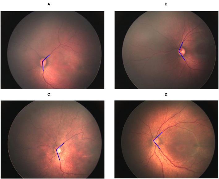
Retinopathy of prematurity (ROP) is a retinal disorder that occurs in preterm infants with low birth weight and is the leading cause of preventable blindness in children. Early identification of high-risk patients and early diagnosis and timely treatment of ROP can substantially improve patients' visual outcomes. However, manual screening consumes both time and resources. Telescreening using retinal fundus images has the potential to reduce the burden engendered by the necessity of on-site screening. Recently, substantial progress has been made in using computer-aided diagnosis with retinal fundus images, and this approach has attracted considerable attention for the diagnosis of eye diseases. Abnormalities of and alterations in retinal blood vessels may relate to the occurrence and progression of ROP. In this study, we examined the hypothesis that ROP severity may be associated with the angle and width of arteries and veins. We computationally determined the artery-artery and vein-vein angles in the temporal quadrants-the temporal artery angle (TAA) and temporal vein angle (TVA)-under normal conditions and in different ROP stages. We also estimated retinal vessel width-temporal artery width (TAW) and temporal vein width (TVW)-by applying the Radon transform method to fundus images. Our results revealed significant decreases in TAA and TVA and increases in TAW and TVW with increasing ROP severity (all P < 0.0001).In addition, we observed positive TAA-TVA and TAW-TVW correlations (both P < 0.0001). The TAA was negatively correlated with the TAW (r = -0.162, P = 0.0314). These retinal vessel features may be useful in assisting ophthalmologists in the early detection of ROP and its progression.
Keywords: Radon transform; computer-aided diagnosis; retinopathy of prematurity (ROP); vessel angle; vessel width.
Copyright © 2022 Huang, Vadloori, Kang and Wu.
Conflict of interest statement
The authors declare that the research was conducted in the absence of any commercial or financial relationships that could be construed as a potential conflict of interest.
Figures


Similar articles
-
Computer-aided detection of retinopathy of prematurity severity assessment via vessel tortuosity measurement in preterm infants' fundus images.Eye (Lond). 2024 Dec;38(17):3309-3317. doi: 10.1038/s41433-024-03285-w. Epub 2024 Aug 3. Eye (Lond). 2024. PMID: 39097674
-
The temporal and nasal retinal arteriolar and venular angles in preterm infants.Br J Ophthalmol. 2011 Dec;95(12):1723-7. doi: 10.1136/bjophthalmol-2011-300416. Epub 2011 Sep 27. Br J Ophthalmol. 2011. PMID: 21951570
-
Computer-aided diagnosis of plus disease via measurement of vessel thickness in retinal fundus images of preterm infants.Comput Biol Med. 2015 Nov 1;66:316-29. doi: 10.1016/j.compbiomed.2015.09.009. Epub 2015 Sep 25. Comput Biol Med. 2015. PMID: 26457930
-
Retinopathy of Prematurity (ROP): An Overview of Biomarkers in Various Samples for Prediction, Diagnosis, and Prognosis.Clin Ophthalmol. 2025 May 7;19:1515-1530. doi: 10.2147/OPTH.S519292. eCollection 2025. Clin Ophthalmol. 2025. PMID: 40357456 Free PMC article. Review.
-
Retinopathy of Prematurity: Therapeutic Strategies Based on Pathophysiology.Neonatology. 2016;109(4):369-76. doi: 10.1159/000444901. Epub 2016 Jun 3. Neonatology. 2016. PMID: 27251645 Review.
Cited by
-
The impact of perinatal brain injury on retinal nerve fiber layer thickness and optic nerve head parameters of premature children.Graefes Arch Clin Exp Ophthalmol. 2023 Sep;261(9):2701-2707. doi: 10.1007/s00417-023-06069-2. Epub 2023 Apr 29. Graefes Arch Clin Exp Ophthalmol. 2023. PMID: 37119306 Free PMC article.
-
Computer-aided detection of retinopathy of prematurity severity assessment via vessel tortuosity measurement in preterm infants' fundus images.Eye (Lond). 2024 Dec;38(17):3309-3317. doi: 10.1038/s41433-024-03285-w. Epub 2024 Aug 3. Eye (Lond). 2024. PMID: 39097674
-
Retinal blood flow velocities in infants with retinopathy of prematurity after intravitreal administration of bevacizumab.Eur J Ophthalmol. 2024 Jan;34(1):95-101. doi: 10.1177/11206721231178062. Epub 2023 May 22. Eur J Ophthalmol. 2024. PMID: 37218176 Free PMC article.
References
LinkOut - more resources
Full Text Sources