. 2022 Jan 28:9:770939.
doi: 10.3389/fcell.2021.770939. eCollection 2021.

Evolution of CDK1 Paralog Specializations in a Lineage With Fast Developing Planktonic Embryos

Affiliations

Evolution of CDK1 Paralog Specializations in a Lineage With Fast Developing Planktonic Embryos

Xiaofei Ma et al. Front Cell Dev Biol. .

Abstract

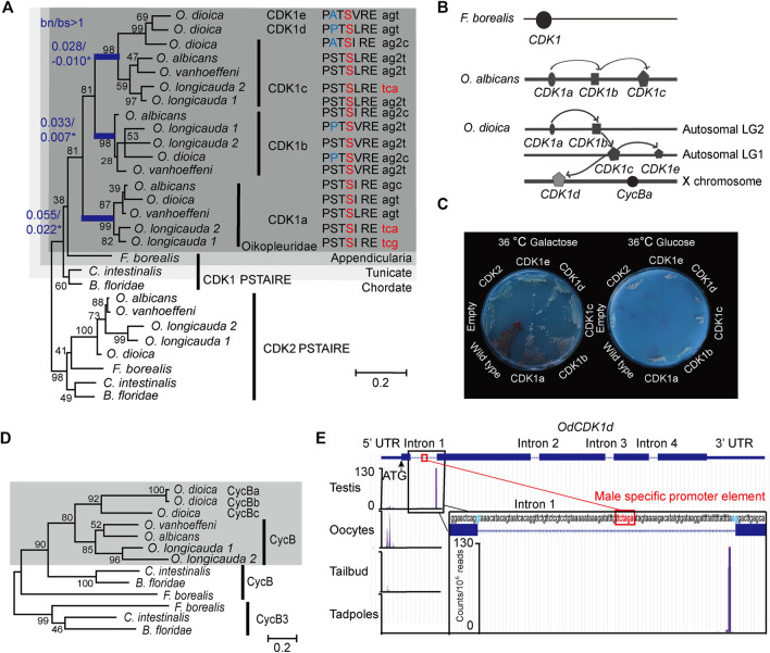
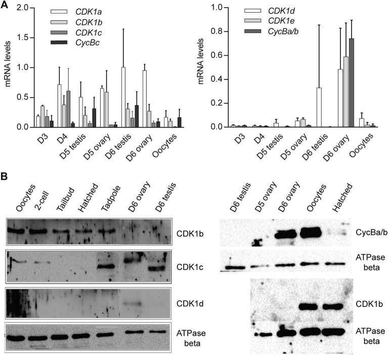
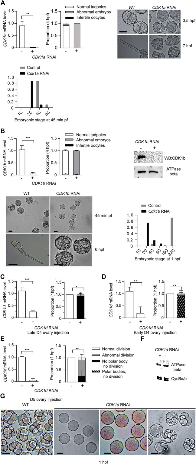
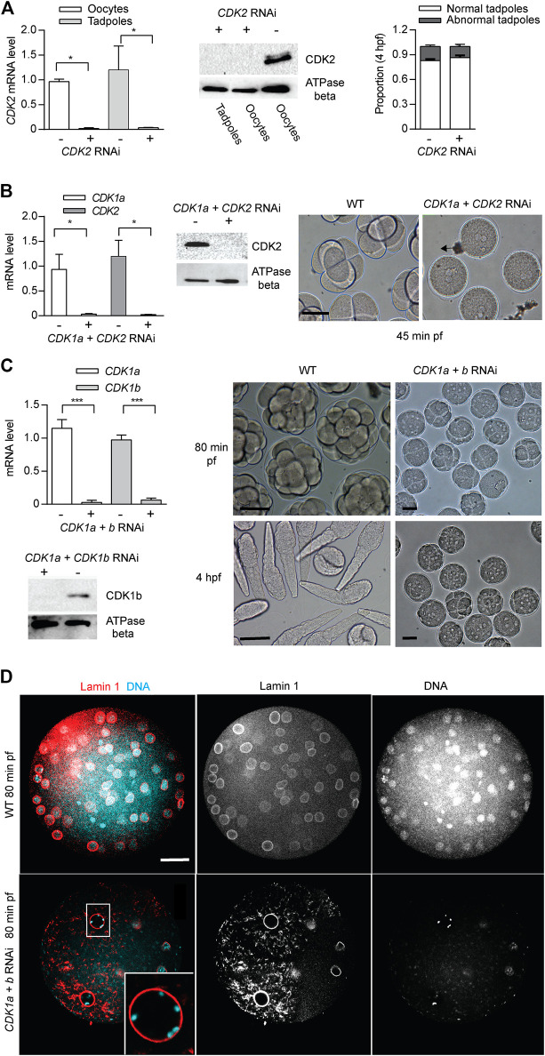
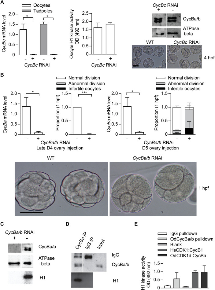
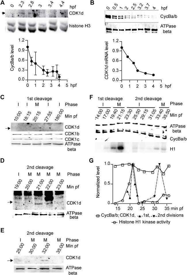
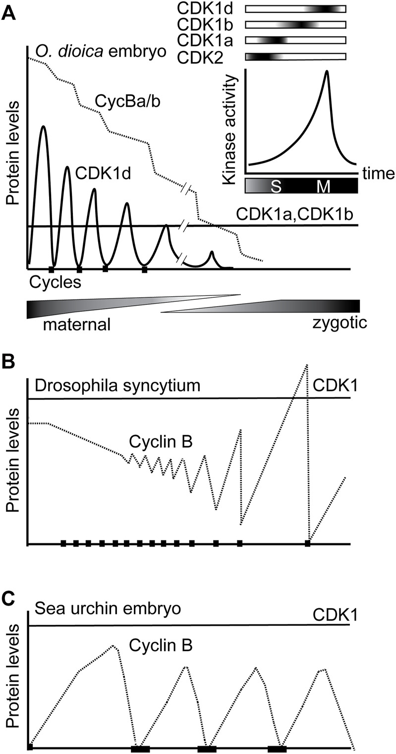
The active site of the essential CDK1 kinase is generated by core structural elements, among which the PSTAIRE motif in the critical αC-helix, is universally conserved in the single CDK1 ortholog of all metazoans. We report serial CDK1 duplications in the chordate, Oikopleura. Paralog diversifications in the PSTAIRE, activation loop substrate binding platform, ATP entrance site, hinge region, and main Cyclin binding interface, have undergone positive selection to subdivide ancestral CDK1 functions along the S-M phase cell cycle axis. Apparent coevolution of an exclusive CDK1d:Cyclin Ba/b pairing is required for oogenic meiosis and early embryogenesis, a period during which, unusually, CDK1d, rather than Cyclin Ba/b levels, oscillate, to drive very rapid cell cycles. Strikingly, the modified PSTAIRE of odCDK1d shows convergence over great evolutionary distance with plant CDKB, and in both cases, these variants exhibit increased specialization to M-phase.

Keywords: PSTAIRE helix; coenocyst; cyclin dependent kinase; determinant of specificity; embryogenesis; gene duplication; nuclear envelope breakdown; tunicate.


Conflict of interest statement

The authors declare that the research was conducted in the absence of any commercial or financial relationships that could be construed as a potential conflict of interest.

Figures

FIGURE 1
FIGURE 1
Amplification of CDK1 paralogs in the Oikopleura genus. (A) Left panel: Maximum likelihood inference analysis of selected chordate CDK1 proteins using the kinase domain of CDK1s with bootstrap values indicated at nodes. bn/bs values in all branches were calculated using a modified Nei-Gojobori method (Zhang et al., 1998). Blue branches have significant bn/bs values over 1 as determined by one-sided Z tests: lineage subtending CDK1a, p = 0.0386; lineage subtending CDK1b, p = 0.0305, lineage subtending CDK1c, p = 0.0334. Modifications in the highly conserved PSTAIRE domain of each CDK1 are shown. The codons resulting in the alanine to serine substitution within the Oikopleuridae (dark grey box) are also indicated. Codons for serine with phase 2 intron insertions (splitting codons between the second and third nucleotides) are denoted by “2”. (B) CDK1 paralog localizations on chromosomes among appendicularians. Arrows indicate possible chronological duplication steps and chromosomal translocations consistent with the phylogenetic analyses. (C) Complementation studies using budding yeast cdc28-4ts mutants substituted with O. dioica CDK1 paralogs. CDK1a, b and d paralogs were able to complement whereas c and e did not. (D) Maximum likelihood inference analysis of selected chordate Cyclin B proteins. The Oikopleuridae paralogs are indicated by a grey box. O. longicauda 1 and 2 indicate two closely related species (or subspecies). (E) Alternative CDK1d transcription start sites (TSS) in testes versus oocytes. CDK1d maternal transcripts have a broad TSS before the ATG start codon. Testes CDK1d transcripts originated from a single TSS immediately before the AG splice acceptor site of fist intron, 42 nt downstream of the TCTAGA male-specific promoter element, which is also located within the first intron. Male CDK1d transcripts lack an ATG start codon, and yield nonsense transcripts in all three possible reading frames. Vertical axis scale for CAGE data (Danks et al., 2018) is in reads per million.
FIGURE 2
FIGURE 2
CDK1 and Cyclin B paralog expression profiles. (A) CDK1-Cyclin B paralog expression levels during gonad maturation from Day 3 to spawning. The data represent means with standard deviations (SD) calculated from independent developmental time courses on three animal populations. Expression data were normalized to transcription of the EF1b and Rpl23 housekeeping genes. Levels are normalized to maximum levels. (B) Western blots of CDK1-Cyclin B paralog levels during development, gonad maturation and embryogenesis. A representative blot from two independent experiments is shown.
FIGURE 3
FIGURE 3
CDK1a, b, and d knockdowns generate abnormal embryonic phenotypes. (A) CDK1a RNAi delays embryonic divisions and tadpoles fail to form. Upper left panel: efficient knockdown of CDK1a in oocytes spawned from ovaries injected with dsRNA against odCDK1a at D5. Upper Mid panel: When exposed to wild-type sperm, CDK1a deficient oocytes generated embryos that divided more slowly than wild-type and failed to hatch at 4 hpf (n1 = 3, n2 = 3). Upper right panel: images of delayed development in CDK1a deficient embryos. When wild-type embryos had developed to the late tailbud stage, CDK1a deficient embryos had only begun to gastrulate. At 7 hpf, tadpoles were observed in wild-type whereas CDK1a deficient embryos failed to reach the tailbud stage. The legend in (A) also applies to the respective panel in (B). Bottom panel: distribution of developmental stages in CDK1a deficient and wild type embryos assessed at 45 min pf. Representative result of three independent experiment is shown. (B) Upper left panel: efficient knockdown CDK1b in oocytes spawned from ovaries injected with dsRNA against odCDK1b at D5 (n1 = 3, n2 = 4). Upper mid panel: When exposed to wild-type sperm, CDK1b deficient oocytes generated embryos that failed to hatch at 4 hpf (n1 = 3, n2 = 4). Upper right panel: Western blot showing absence of CDK1b protein in CDK1b RNAi embryos. Representative blot from three independent experiments is shown. Bottom left panel: CDK1b deficient embryos arrested before gastrulation (n1 = 3, n2 = 3). Bottom right panel: distribution of developmental stages in CDK1b deficient and wild type embryos assessed at 1 hpf. Representative result of three independent experiment is shown. (C–E), Knockdown of CDK1d generated 3 different phenotypes at 1 hpf. (C) Left panel: efficient knockdown in oocytes spawned from ovaries that had been injected with dsRNA against odCDK1d at late D4 (C) (n1 = 3, n2 = 3). Right panel: abnormal division in all cases with a subset of embryos exhibiting more severe phenotypes of polar body extrusion but no cell division, or a complete lack of polar body extrusion and absence of any division (n1 = 3, n2 = 3). (D) Left panel: efficient knockdown in oocytes spawned from ovaries that had been injected with dsRNA against odCDK1d at early D5 (n1 = 3, n2 = 3). Right panel: proportion of embryonic phenotypes for CDK1d deficient embryos (n1 = 3, n2 = 3). (E) Left panel: efficient knockdown in oocytes spawned from ovaries that had been injected with dsRNA against odCDK1d at D5 (n1 = 3, n2 = 3). Right panel: proportion of embryonic phenotypes for CDK1d deficient embryos (n1 = 3, n2 = 3). The legend in (E) also applies to the respective panels in (C) and (D). (F) Knockdown of CDK1d did not have any effect on the levels of Cyclin Ba/b, the Cyclin component of MPF in O. dioica oocytes (Feng and Thompson 2018). Representative blot from two independent experiments is shown. (G) Image comparisons of wild-type to CDK1d knockdown oocyte/embryo phenotypes 1 h post exposure to wild-type sperm. Left to right: WT cleavage stage embryos, infertile oocytes with no polar body extrusion, polar bodies extruded, but no division, and abnormally dividing embryos. Where indicated, data are mean (SD); ***p < 0.001, **p < 0.01, *p < 0.05. Scale bars: 50 µm.
FIGURE 4
FIGURE 4
CDK2 is dispensable for Oikopleura dioica embryogenesis. Synergistic defects of CDK2 + CDK1a RNAi and CDK2 + CDK1b RNAi in embryonic divisions. (A) CDK2 is dispensable for embryonic divisions. Left panel: efficient knockdown of CDK2 in oocytes and tadpoles, spawned from ovaries injected with dsRNA against odCDK2 at D4 (n1 = 3, n2 = 3). Mid panel: Western blot showing absence of CDK2 protein in CDK2 RNAi oocytes and tadpoles. Representative blot from two independent experiments is shown. Right panel: no significant differences in developmental outcome between CDK2 deficient and wild-type embryos (n1 = 3, n2 = 3). (B) Synergistic effect of CDK2 + CDK1a RNAi on embryonic divisions. Left Panel: efficient knockdown of CDK1a and CDK2 in oocytes from ovaries that had been injected with dsRNA against odCDK2 and CDK1a at D5 (n1 = 3, n2 = 3). Mid panel: Western blot showing absence of CDK2 protein in CDK1a + CDK2 RNAi in oocytes. Representative blot from three independent experiments is shown. Right panel: at 45 min pf, CDK1a + CDK2 deficient zygotes were arrested prior to pronuclear fusion. Scale bars: 50 µm. (C) Synergistic effect of CDK1a + b RNAi on embryonic division. Upper left panel: efficient knockdown of CDK1a and b in oocytes from ovaries that had been injected with dsRNA against CDK1a + b at D5 (n1 = 3, n2 = 3). Lower left: Western blot showing absence of CDK1b protein in CDK1a + b RNAi oocytes. Representative blot from three independent experiments is shown. Right panel: CDK1a + b deficient zygotes finished 3 rounds of slowed divisions before arresting at the 4th–5th embryonic division. Scale bars: 50 µm. Where indicated, date are mean (SD); ***p < 0.001, **p < 0.01, *p < 0.05. (D) Confocal images of Lamin 1 and DNA staining of wild-type versus CDK1a + b RNAi embryos at 80 min post-fertilization. CDK1a + b deficient zygotes arrest at prophase with super-condensed chromosomes at the nuclear periphery (inset) after the third division. Scale bars: 20 µm.
FIGURE 5
FIGURE 5
Cyclin Ba/b, is required for O. dioica embryonic cell cycles, whereas Cyclin Bc is dispensable. (A) Cyclin Bc is dispensable for embryonic divisions. Left panel: efficient knockdown cycBc in oocytes (n1 = 3, n2 = 3) and tadpoles (n1 = 2, n2 = 3) spawned from ovaries that had been injected with dsRNA against cycBc at D5. Mid panel: no significant difference in mean H1 kinase activity between cycBc deficient and wild-type oocytes (metaphase I arrested, n1 = 3, n2 = 3). Upper right panel: Western blot showing that knockdown of cycBc had no effect on protein levels of Cyclin Ba/b. Representative blot from three independent experiments is shown. Lower right panel: images show Cyclin Bc deficient and wild-type embryos had identical phenotypes at the tailbud stage, 4 hpf. (B) Knockdown of cycBa/b generated 2 different phenotypes at 1 hpf. Upper 1st panel: efficient knockdown of cycBa/b in oocytes spawned from ovaries that had been injected with dsRNA against cycBa/b at late D4 (n1 = 3, n2 = 3). Upper 2nd panel: Cyclin Ba/b deficient oocytes exposed to wild type sperm generated two embryonic phenotypes: infertile and abnormal divisions. Data represent the proportion of phenotypes in 50 zygotes derived from each ovary (n1 = 3, n2 = 3). Upper 3rd panel: efficient knockdown of cycBa/b in oocytes spawned from ovaries that had been injected with dsRNA against cycBa/b at D5 (n1 = 3, n2 = 3). Upper 4th panel: proportion of embryonic phenotypes for Cyclin Ba/b deficient oocytes spawned form ovaries that had been injected with dsRNA against cycBa/b at D5. Where indicated, data are mean (SD); ***p < 0.001, **p < 0.01, *p < 0.05. Bottom panel: images show abnormal division phenotypes, compared to wild-type at 1 hpf. Scale bars: 50 µm. (C–E) CycBa/b-CDK1d complexes have histone H1 kinase activity. (C) H1 kinase activity was present in wild type oocytes but absent in cycBa/b RNAi knockdown oocytes (Feng and Thompson, 2018). (D) Cyclin Ba/b immunoprecipitates from oocytes had H1 kinase activity whereas IgG oocyte immunoprecipitates did not. Representative result from three independent experiments is shown. (E) H1 kinase activity levels of O. dioica Cyclin Ba/b pulldowns compared to IgG pulldowns and in vitro expressed complexes of Human (Hs) CDK1:Cyclin B1 and O. dioica (Od) CDK1d:Cyclin Ba. The data represent mean (SD) of three independent experiment.
FIGURE 6
FIGURE 6
CDK1d levels oscillate during early embryonic cell cycles. (A) Upper panel: Western blot of CDK1d levels during the first 4 h of development with histone H3 as a loading control. Lower panel: CDK1d mRNA levels were quantified during early embryonic development by qRT-PCR, normalized to levels in unfertilized oocytes. (B) Cyclin Ba/b levels do not oscillate but instead, decline steadily during early embryogenesis. Upper panel: Western blot of Cyclin Ba/b during the first 4 h of development with ATPase beta as a loading control. Lower panel: quantification of Cyclin Ba/b levels, normalized to levels in unfertilized oocytes. The data represent mean (SD) of three independent experiments. (C–E) CDK1d levels peaked in M phase of first two mitotic embryonic cell cycles. (C) Western blot of CDK1b, c, and d levels during the first 3 hpf. Arrow indicates the CDK1d band. I, interphase; M, M-phase. (D) Sampling of CDK1d levels at 1-min internals around M phase of the first embryonic cell cycle. Arrow indicates the oscillation of the CDK1d band. (E) Sampling of CDK1d levels around M-phase of the second division cycle. Arrow indicates the oscillation of the CDK1d band. (F) Western blot analyses of Cyclin Ba/b levels and histone H1 kinase activity during the first two embryonic cell cycles. Cyclin Ba/b levels remained elevated, whereas H1 kinase activity oscillated, with peaks in the first two M-phases. Representative result from three independent experiments is shown. (G) Quantification of Cyclin Ba/b, CDK1d and H1 kinase activity during the first two embryonic cell cycles.
FIGURE 7
FIGURE 7
Sequential activation of CDK1a, b and d, and switching from oscillations of CDK1d levels to oscillation of Cyclin Ba/b levels, drive rapid early embryonic cell cycles in O. dioica. (A) Knockdowns revealed that sequential activation of 3 O. dioica CDK1 paralogs, a, b and d, are required for proper execution of embryonic divisions leading to embryo hatching. Cyclin Ba/b, the partner of CDK1d, is maternally stocked and gradually degrades over the first embryonic cycles, whereas unusually, it is CDK1d protein levels that initially oscillate to regulate rapid M-phases. CDK1a and b levels remain constant during this period. During the maternal to zygotic transition, the cell cycle slows and divisions become less synchronous. In the coenocystic ovary, polyploid nurse nuclei are afforded the time to produce maternally stocked transcripts, many arising from genes containing multiple introns of variable length, with the genes often organized in polycistronic operons (Danks et al., 2015). During the period of rapid embryonic cell divisions, many zygotically activated genes are monocistronic, and contain short introns, or are intronless. (B) Modified from Edgar et al. (1994). Due to excess Cyclin B protein storage in Drosophila embryos, there is little fluctuation in Cyclin B levels during cycles 1–7. Drosophila syncytial embryos have adapted Cyclin B preloading to mitosis without cytokinesis, compatible with little oscillation of CDK1 kinase levels due to localized Cyclin B destruction. Beginning at cycle 8, cyclin fluctuation becomes apparent. (C) As in sea urchin embryos, it is widely conserved that CDK1 protein levels are constant while Cyclin B levels, oscillate, across cell cycles.

Similar articles

Cited by

References

    1. Ahn S.-H., Tobe B. T., Gerald J. N. F., Anderson S. L., Acurio A., Kron S. J. (2001). Enhanced Cell Polarity in Mutants of the Budding Yeast Cyclin-dependent Kinase Cdc28p. MBoC 12, 3589–3600. 10.1091/mbc.12.11.3589 - DOI - PMC - PubMed
    1. Arooz T., Yam C. H., Siu W. Y., Lau A., Li K. K. W., Poon R. Y. C. (2000). On the Concentrations of Cyclins and Cyclin-dependent Kinases in Extracts of Cultured Human Cells. Biochemistry 39, 9494–9501. 10.1021/bi0009643 - DOI - PubMed
    1. Bachtrog D., Jensen J. D., Zhang Z. (2009). Accelerated Adaptive Evolution on a Newly Formed X Chromosome. Plos Biol. 7, e28e1000082. 10.1371/journal.pbio.1000082 - DOI - PMC - PubMed
    1. Bao Z. Q., Jacobsen D. M., Young M. A. (2011). Briefly Bound to Activate: Transient Binding of a Second Catalytic Magnesium Activates the Structure and Dynamics of Cdk2 Kinase for Catalysis. Structure 19, 675–690. 10.1016/j.str.2011.02.016 - DOI - PMC - PubMed
    1. Bentley A. M., Normand G., Hoyt J., King R. W. (2007). Distinct Sequence Elements of Cyclin B1 Promote Localization to Chromatin, Centrosomes, and Kinetochores during Mitosis. MBoC 18, 4847–4858. 10.1091/mbc.E06-06-0539 - DOI - PMC - PubMed