Review
. 2022 Jan 26:9:801652.
doi: 10.3389/fcell.2021.801652. eCollection 2021.

Turning the Curve Into Straight: Phenogenetics of the Spine Morphology and Coordinate Maintenance in the Zebrafish

Affiliations
Review

Turning the Curve Into Straight: Phenogenetics of the Spine Morphology and Coordinate Maintenance in the Zebrafish

Carlos Muñoz-Montecinos et al. Front Cell Dev Biol. .

Abstract

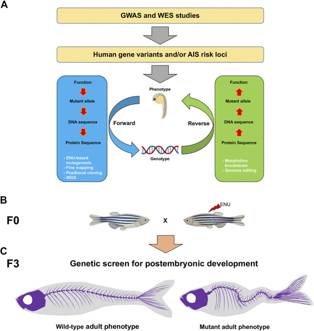
The vertebral column, or spine, provides mechanical support and determines body axis posture and motion. The most common malformation altering spine morphology and function is adolescent idiopathic scoliosis (AIS), a three-dimensional spinal deformity that affects approximately 4% of the population worldwide. Due to AIS genetic heterogenicity and the lack of suitable animal models for its study, the etiology of this condition remains unclear, thus limiting treatment options. We here review current advances in zebrafish phenogenetics concerning AIS-like models and highlight the recently discovered biological processes leading to spine malformations. First, we focus on gene functions and phenotypes controlling critical aspects of postembryonic aspects that prime in spine architecture development and straightening. Second, we summarize how primary cilia assembly and biomechanical stimulus transduction, cerebrospinal fluid components and flow driven by motile cilia have been implicated in the pathogenesis of AIS-like phenotypes. Third, we highlight the inflammatory responses associated with scoliosis. We finally discuss recent innovations and methodologies for morphometrically characterize and analyze the zebrafish spine. Ongoing phenotyping projects are expected to identify novel and unprecedented postembryonic gene functions controlling spine morphology and mutant models of AIS. Importantly, imaging and gene editing technologies are allowing deep phenotyping studies in the zebrafish, opening new experimental paradigms in the morphometric and three-dimensional assessment of spinal malformations. In the future, fully elucidating the phenogenetic underpinnings of AIS etiology in zebrafish and humans will undoubtedly lead to innovative pharmacological treatments against spinal deformities.

Keywords: CSF-cNs; Reissner fiber; cerebrospinal fluid; cilia; inflammation; scoliosis; spine; zebrafish.


Conflict of interest statement

The authors declare that the research was conducted in the absence of any commercial or financial relationships that could be construed as a potential conflict of interest.

Figures

FIGURE 1
FIGURE 1
Schematic of the genotype-phenotype association and genetic approaches to study AIS candidate genes in the zebrafish. (A) To identify AIS risk loci, GWAS and WES strategies can be performed. Forward (blue) and reverse (green) genetic strategies (white) are powerful tools to study mutant genotypes and phenotypes associated with AIS. (B) In a forward genetic screen, male founders (F0) are mutagenized with ENU and then outcrossed to generate F2 families. To screen for postembryonic gene functions in late larval to adult stages after 5 dpf, F3 families are generated either through incrossing F2 heterozygous fish or through in vitro fertilization (IVF) using cryopreserved sperm and wild-type females, and raised to adulthood. (C) By using a combination of morphological, tomographic, molecular and cellular approaches, F3 individuals are screened for adult spine alterations. AIS, adolescent idiopathic scoliosis; GWAS, genome-wide association studies; WES, whole exome sequencing; ENU, N-ethyl-N-nitrosourea; WGS, whole genome sequencing.
FIGURE 2
FIGURE 2
Schematic representation of the proprioceptive organ conformed by RF and CSF-cNs. (A) The central canal in a zebrafish embryo is surrounded by ependymal ciliated cells (gray) necessary for the CSF flow, and by sensory CSF-cNs (yellow). The RF (green cord) runs from the SCO to the entire spinal cord inside the central canal. The RF binds and transports epinephrine and norepinephrine molecules (red dots) that trigger Urp1 and Urp2 expression in mechanosensory CSF-cNs. (B) The curvature of the spinal cord during locomotion, allows the interaction between RF and CSF-cNs (highlighted in red); thus, transferring information through a spinal circuit and shaping the spinal curvature. (C) CSF-cNs project an ipsilateral ascending axon that synapses onto premotor INs, onto primary MNs or directly onto muscle that expresses the Urp receptor. Additionally, the most cephalic CSF-cNs send their axons to RSNs in the hindbrain, that in turn send descending motor commands to the spinal cord. All these projections participate in the correct muscle contraction and postural control. SCO, subcommissural organ; CSF, cerebrospinal fluid; CSF-cNs, CSF contacting neurons; RF, Reissner fiber; Urp, urotensin neuropeptides; IN, interneuron; MN, motor neuron; RSN, reticulospinal neuron.
FIGURE 3
FIGURE 3
Schematic representation of macrophages activation and spatial distribution, and the regionalized inflammatory response triggered during scoliosis development. Colored macrophages (purple) are shown. At the onset of the spinal curve formation, immature macrophages are distributed along the ventral side of the tail. After their activation, these cells move and accumulate within the spinal cord. Macrophages maturation also triggers a neuroinflammatory response associated with TNF-α overexpression in the brain. TNF-α, tumor necrosis factor alpha.

References

    1. Acaroglu R. E., Akel A., Alanay M., Yazici M., Marcucio R. (2009). Comparison of the melatonin and calmodulin in paravertebral muscle and platelets of patients with or without adolescent ıdiopathic scoliosis. Spine J. 18, E659-63. - PubMed
    1. Adham I. M., Gille M., Gamel A. J., Reis A., Dressel R., Steding G., et al. (2005). The Scoliosis (Sco) Mouse: a New Allele of Pax1. Cytogenet. Genome Res. 111 (1), 16–26. 10.1159/000085665 - DOI - PubMed
    1. Alejevski F., Leemans M., Gaillard A.-L., Leistenschneider D., de Flori C., Bougerol M., et al. (2021). Conserved Role of the Urotensin II Receptor 4 Signalling Pathway to Control Body Straightness in a Tetrapod. Open Biol. 11 (8), 210065. 10.1098/rsob.210065 - DOI - PMC - PubMed
    1. Algin O., Koc U., Yalcin N. (2021). Cerebrospinal Fluid Velocity Changes of Idiopathic Scoliosis: a Preliminary Study on 3-T PC-MRI and 3D-SPACE-VFAM Data. Childs Nerv Syst. 10.1007/s00381-021-05339-w - DOI - PubMed
    1. Andersen M. O., Thomsen K., Kyvik K. O. (20071976). Adolescent Idiopathic Scoliosis in Twins. Spine 32 (8), 927–930. 10.1097/01.brs.0000259865.08984.00 - DOI - PubMed

LinkOut - more resources