. 2022 Jan 28:8:830730.
doi: 10.3389/fmed.2021.830730. eCollection 2021.

Diagnostic Accuracy of H. pylori Status by Conventional Endoscopy: Time-Trend Change After Eradication and Impact of Endoscopic Image Quality

Affiliations

Diagnostic Accuracy of H. pylori Status by Conventional Endoscopy: Time-Trend Change After Eradication and Impact of Endoscopic Image Quality

Duc Trong Quach et al. Front Med (Lausanne). .

Abstract

Aim: To assess the time trend of diagnostic accuracy of pre- and post-eradication H. pylori status and interobserver agreement of gastric atrophy grading.

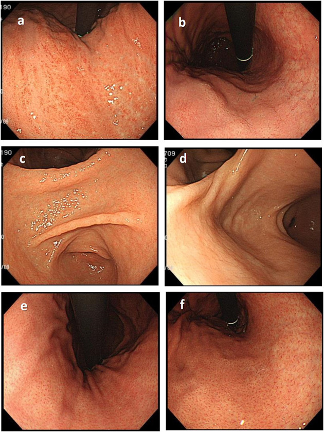
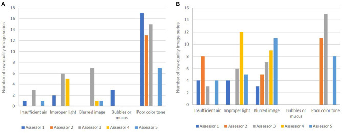
Methods: A series 100 of conventional endoscopic image sets taken from subjects undergoing gastric cancer screening at a polyclinic were evaluated by 5 experienced assessors. Each assessor independently examined endoscopic findings according to the Kyoto classification and then determined the H. pylori status (never, current, or past infected). Gastric atrophy was assessed according to the Kimura-Takemoto classification and classified into 3 grades (none/mild, moderate, or severe). The image series that ≥3 assessors considered to have good quality were arbitrarily defined as high-quality image (HQI) series, and the rest were defined as low-quality image (LQI) series.

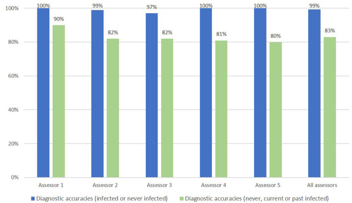
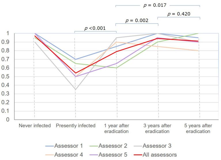
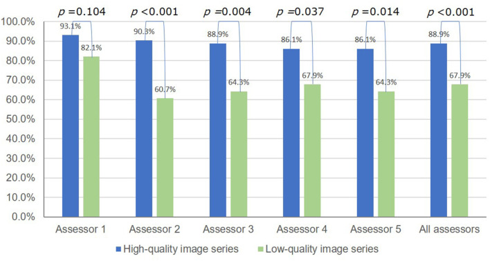
Results: The overall diagnostic accuracy of H. pylori status was 83.0%. It was lowest in subjects with current infection (54%), gradually increased at 1 year (79%, P < 0.001) and 3 years (94.0%, P = 0.002), but then did not significantly change at 5 years (91.0%, P = 0.420) after eradication. The rate of LQI series was 28%. The overall diagnostic accuracy of H. pylori status dropped from 88.9% to 67.9% (P < 0.001), and the mean kappa value on gastric atrophy grading dropped from 0.730 to 0.580 (P = 0.002) in the HQI and LQI series, respectively.

Conclusions: Diagnostic accuracy of H. pylori status increased over time after eradication. LQI series badly affected the diagnostic accuracy of H. pylori status and the level of agreement when grading gastric atrophy.

Keywords: Helicobacter pylori; Kimura-Takemoto classification; Kyoto classification; endoscopic diagnosis; gastric atrophy; interobserver agreement.


Conflict of interest statement

The authors declare that the research was conducted in the absence of any commercial or financial relationships that could be construed as a potential conflict of interest.

Figures

Figure 1
Figure 1
Important endoscopic findings and pitfalls in the correct diagnosis of H. pylori status. (a) Diffuse redness, typically seen in patients with current H. pylori infection, should be differentiated from patchy redness (b), which can be seen in patients with past infection. (c) Mucosal edema is a typical finding commonly seen in current H. pylori infection that disappears in patients with past infection (d). The regular arrangement of collecting venules (e), which is a typical finding in the stomach of patients never infected with H. pylori, disappears in patients with H. pylori infection but could recur years after eradication (f).
Figure 2
Figure 2
Endoscopic accuracy of H. pylori infection diagnosis (infected/never infected) and accuracy for H. pylori infection status (never, current, or past infected).
Figure 3
Figure 3
Time trend of diagnostic accuracy for H. pylori status based on white-light endoscopy.
Figure 4
Figure 4
Accuracy of the endoscopic diagnosis of H. pylori infection status (never, current, or past infected) using high-quality vs. low-quality image series.
Figure 5
Figure 5
Causes of low-quality images used for the endoscopic diagnosis of H. pylori status (A) and for gastric atrophy grading (B).
Figure 6
Figure 6
Examples of low-quality endoscopic images. (a) Insufficient air insufflation (image obtained in a patient at 4 years after eradication). (b) Improper light (too dark) (image obtained in a patient with current H. pylori infection). (c) Improper light (too bright) (image obtained in a patient with current H. pylori infection). (d) Blurred image (image obtained in a patient with current H. pylori infection). (e) Some mucus is still retained in the stomach (image obtained in a patient at 5 years after eradication). (f) Poor color tone (image obtained in a patient at 1 year after eradication).

Similar articles

References

    1. Bang CS, Lee JJ, Baik GH. Artificial intelligence for the prediction of helicobacter pylori infection in endoscopic images: systematic review and meta-analysis of diagnostic test accuracy. J Med Internet Res. (2020) 22:e21983. 10.2196/21983 - DOI - PMC - PubMed
    1. Chiu PWY, Uedo N, Singh R, Gotoda T, Ng EKW, Yao K, et al. . An Asian consensus on standards of diagnostic upper endoscopy for neoplasia. Gut. (2019) 68:186–97. 10.1136/gutjnl-2018-317111 - DOI - PubMed
    1. Dohi O, Majima A, Naito Y, Yoshida T, Ishida T, Azuma Y, et al. . Can image-enhanced endoscopy improve the diagnosis of Kyoto classification of gastritis in the clinical setting? Digestive Endoscopy. (2019) 32:191–203. 10.1111/den.13540 - DOI - PubMed
    1. Glover B, Teare J, Ashrafian H, Patel N. The endoscopic predictors of Helicobacter pylori status: a meta-analysis of diagnostic performance. Therapeutic Adv Gastrointest Endosc. (2020) 13:2631774520950840. 10.1177/2631774520950840 - DOI - PMC - PubMed
    1. Hiyama T, Quach DT, Le QD, Ho LX, Vu NHT, Shimamoto F, et al. . Rate of Unintended Helicobacter pylori Eradication in the Vietnamese. Helicobacter. (2015) 20:156–7. 10.1111/hel.12210 - DOI - PubMed