. 2021 Oct:1:732243.
doi: 10.3389/fnetp.2021.732243. Epub 2021 Oct 12.

Circadian Synchrony: Sleep, Nutrition, and Physical Activity

Affiliations

Circadian Synchrony: Sleep, Nutrition, and Physical Activity

Kelly L Healy et al. Front Netw Physiol. 2021 Oct.

Abstract

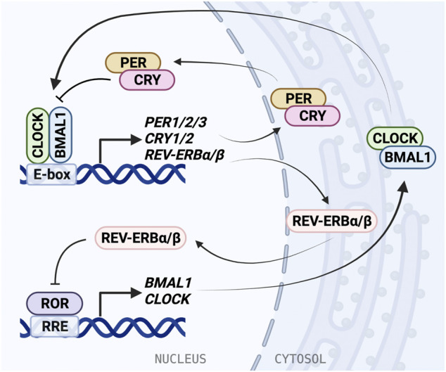
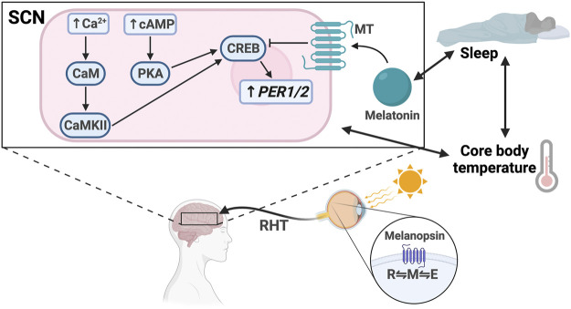
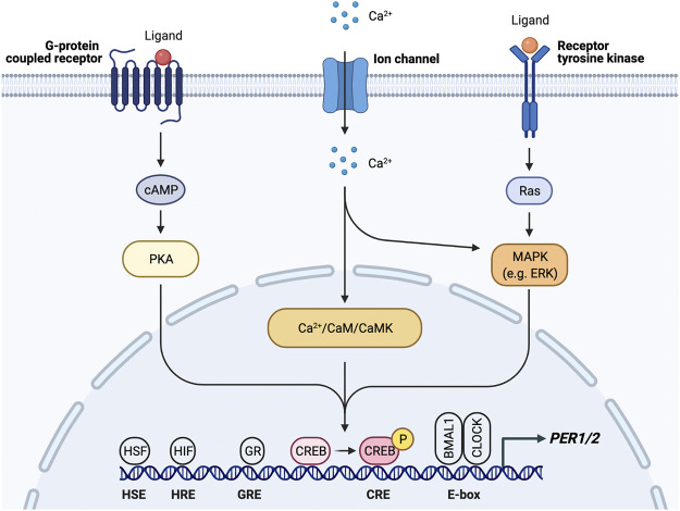
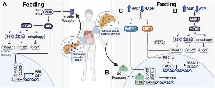
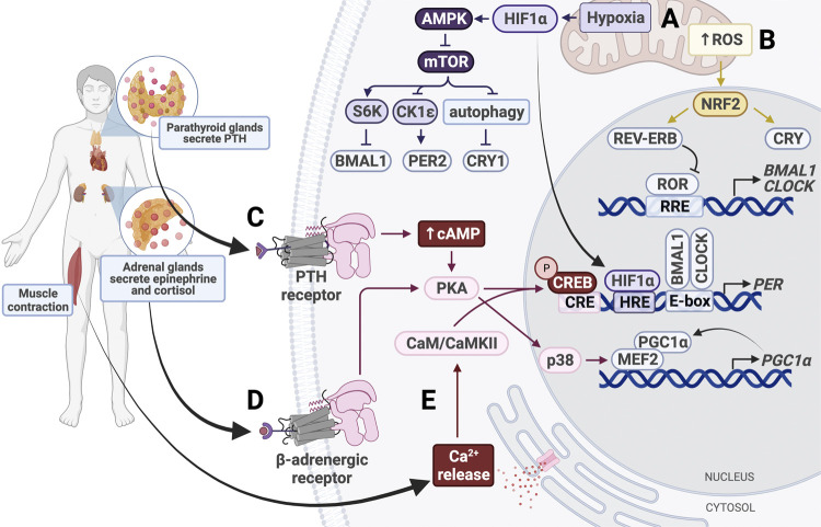
The circadian clock in mammals regulates the sleep/wake cycle and many associated behavioral and physiological processes. The cellular clock mechanism involves a transcriptional negative feedback loop that gives rise to circadian rhythms in gene expression with an approximately 24-h periodicity. To maintain system robustness, clocks throughout the body must be synchronized and their functions coordinated. In mammals, the master clock is located in the suprachiasmatic nucleus (SCN) of the hypothalamus. The SCN is entrained to the light/dark cycle through photic signal transduction and subsequent induction of core clock gene expression. The SCN in turn relays the time-of-day information to clocks in peripheral tissues. While the SCN is highly responsive to photic cues, peripheral clocks are more sensitive to non-photic resetting cues such as nutrients, body temperature, and neuroendocrine hormones. For example, feeding/fasting and physical activity can entrain peripheral clocks through signaling pathways and subsequent regulation of core clock genes and proteins. As such, timing of food intake and physical activity matters. In an ideal world, the sleep/wake and feeding/fasting cycles are synchronized to the light/dark cycle. However, asynchronous environmental cues, such as those experienced by shift workers and frequent travelers, often lead to misalignment between the master and peripheral clocks. Emerging evidence suggests that the resulting circadian disruption is associated with various diseases and chronic conditions that cause further circadian desynchrony and accelerate disease progression. In this review, we discuss how sleep, nutrition, and physical activity synchronize circadian clocks and how chronomedicine may offer novel strategies for disease intervention.

Keywords: asynchrony; circadian misalignment; circadian rhythm; entrainment; synchronization.


Conflict of interest statement

Conflict of Interest: The authors declare that the research was conducted in the absence of any commercial or financial relationships that could be construed as a potential conflict of interest.

Figures

FIGURE 1
FIGURE 1
The molecular mechanism of the mammalian circadian clock. The molecular clock is based on a transcriptional/translational negative feedback mechanism and consists of the core loop and two interlocking loops. In the core loop, the BMAL1/CLOCK heterodimer binds to the E-box cis-elements and drives transcription of targeted genes, including PER1/2/3, CRY1/2, and NR1D1/2 (REV-ERBα/β). PER and CRY inhibit BMAL1/CLOCK activity and therefore repress their own expression. In the RRE loop, REV-ERB repressors and ROR activators bind to RRE to regulate BMAL1 and CLOCK expression.
FIGURE 2
FIGURE 2
Light entrainment of the SCN and sleep/wake regulation. Light strikes the retina and excites melanopsin, driving it to the M configuration. The signal travels along the retinohypothalamic tract (RHT), resulting in increased intracellular levels of Ca2+ and cAMP in the SCN. Ca2+ activates calmodulin (CaM) and CaMKII. cAMP activates PKA. CaMKII and PKA activate CREB, which drives PER1/2 transcription. Melatonin binds the melatonin receptor (MT), which inhibits CREB activation. There is bidirectional regulation between melatonin secretion and sleep, sleep and core body temperature, and core body temperature and SCN signaling.
FIGURE 3
FIGURE 3
PER1/2 transcriptional regulation by multiple upstream signals. Ca2+/cAMP signaling cascade activates CREB by phosphorylation. Activated CREB binds the cAMP response element (CRE) to drive PER1/2 transcription. Other proteins (HSF, heat shock factor; HIF, hypoxia inducible factor; GR, glucocorticoid receptor) recognize and bind other consensus sequences (HSE, heat shock element; HRE, hypoxia response element; GRE, glucocorticoid response element).
FIGURE 4
FIGURE 4
Entrainment of peripheral clocks by feeding/fasting. (A) After feeding, insulin activates PI3K/AKT, which inhibits BMAL1 and activates mTOR. (B) During fasting, cortisol binds the glucocorticoid (GC) receptor, which binds glucocorticoid response elements (GREs) in promoters to drive PER1/2 transcription. (C) Also during fasting, PARP-1 and SIRT1 are activated in response to an increased ratio of NAD+/NADH. Subsequent post-translational modifications alter expression of core clock genes. (D) Higher ratio of AMP/ATP activates the AMPK, a mTOR inhibitor.
FIGURE 5
FIGURE 5
Entrainment of peripheral clocks by physical activity. (A) Hypoxic conditions activate HIF1α and AMPK, which inhibits mTOR activity. HIF1α binds the HIF response element (HRE) in PER1/2 promoters to increase gene expression. (B) Increased intracellular levels of reactive oxygen species (ROS) activate NRF2, which subsequently activates CRY and REV-ERB. (C) Parathyroid hormone (PTH) binds the PTH receptor to activate the cAMP/Ca2+-CREB signaling cascade. Phosphorylated CREB binds the cAMP response element (CRE) in the PER promoter, thereby upregulating PER expression. (D) Secreted epinephrine binds the β-adrenergic receptor, which activates protein kinase A (PKA). PKA activates CREB and p38 to alter clock gene expression. p38 triggers activation of the MEF2/PGC1α positive feedback loop. (E) Muscle contraction triggers intracellular calcium release, which activates calmodulin (CaM) and calmodulin-dependent kinase II (CaMKII). CaM/CaMKII activate CREB, which upregulates PER1/2 expression.

References

    1. Abou-Samra A. B., Jüppner H., Force T., Freeman M. W., Kong X. F., Schipani E., et al. (1992). Expression Cloning of a Common Receptor for Parathyroid Hormone and Parathyroid Hormone-Related Peptide from Rat Osteoblast-like Cells: a Single Receptor Stimulates Intracellular Accumulation of Both cAMP and Inositol Trisphosphates and Increases Intracellular Free Calcium. Proc. Natl. Acad. Sci. 89, 2732–2736. 10.1073/pnas.89.7.2732 - DOI - PMC - PubMed
    1. Adafer R., Messaadi W., Meddahi M., Patey A., Haderbache A., Bayen S., et al. (2020). Food Timing, Circadian Rhythm and Chrononutrition: A Systematic Review of Time-Restricted Eating's Effects on Human Health. Nutrients 12, 3770. 10.3390/nu12123770 - DOI - PMC - PubMed
    1. Aghaloo T. L., Pirih F. Q., Shi A., Bezouglaia O., Tetradis S. (2006). Parathyroid Hormone Induces Mitogen-Activated Kinase Phosphatase 1 in Murine Osteoblasts Primarily through cAMP-Protein Kinase A Signaling. J. Periodontol. 77, 21–30. 10.1902/jop.2006.77.1.21 - DOI - PubMed
    1. Akashi M., Tsuchiya Y., Yoshino T., Nishida E. (2002). Control of Intracellular Dynamics of Mammalian Period Proteins by Casein Kinase I ε (CKIε) and CKIδ in Cultured Cells. Mol. Cel Biol 22, 1693–1703. 10.1128/MCB.22.6.1693-1703.2002 - DOI - PMC - PubMed
    1. Akimoto T., Pohnert S. C., Li P., Zhang M., Gumbs C., Rosenberg P. B., et al. (2005). Exercise Stimulates Pgc-1α Transcription in Skeletal Muscle through Activation of the P38 MAPK Pathway. J. Biol. Chem. 280, 19587–19593. 10.1074/jbc.M408862200 - DOI - PubMed