. 2022 Apr;9(11):e2105059.
doi: 10.1002/advs.202105059. Epub 2022 Feb 13.

Arrested in Glass: Actin within Sophisticated Architectures of Biosilica in Sponges

Affiliations

Arrested in Glass: Actin within Sophisticated Architectures of Biosilica in Sponges

Hermann Ehrlich et al. Adv Sci (Weinh). 2022 Apr.

Abstract

Actin is a fundamental member of an ancient superfamily of structural intracellular proteins and plays a crucial role in cytoskeleton dynamics, ciliogenesis, phagocytosis, and force generation in both prokaryotes and eukaryotes. It is shown that actin has another function in metazoans: patterning biosilica deposition, a role that has spanned over 500 million years. Species of glass sponges (Hexactinellida) and demosponges (Demospongiae), representatives of the first metazoans, with a broad diversity of skeletal structures with hierarchical architecture unchanged since the late Precambrian, are studied. By etching their skeletons, organic templates dominated by individual F-actin filaments, including branched fibers and the longest, thickest actin fiber bundles ever reported, are isolated. It is proposed that these actin-rich filaments are not the primary site of biosilicification, but this highly sophisticated and multi-scale form of biomineralization in metazoans is ptterned.

Keywords: actin; biological materials; biomineralization; biosilica; sponges.


Conflict of interest statement

The authors declare no conflict of interest.

Figures
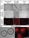
Figure 1
Figure 1
Identification of actin in the axial filaments of diverse sponge species using iFluor 594‐Phalloidin red stain. a) Overview of stained axial filaments isolated from selected biosilica‐based structures such as tauactines of M. chuni (Hexactinellida), megascleres (oxea) of S. lacustris (Demospongiae), and skeletal framework of Farrea. sp. (Hexactinellida) following 10% HF treatment. All the axial filaments, isolated from both marine (M. chuni, Farrea sp.) and freshwater (S. lacustris) sponges, resemble the size and morphology of the siliceous skeletal structures they were derived from. b) HF‐based treatment of siliceous spherical microspined rays forming the oxyasters of G. cydonium marine demosponge led to the isolation of an organic matrix with branching, radially spaced microfibers also visible after iFluor 594‐Phalloidin staining (see also Figure S10, Supporting Information).
Figure 2
Figure 2
Confirmation of the presence of actin in axial filaments of selected sponges species using immunofluorescent analysis with primary anti‐β‐actin antibody and secondary (anti‐rabbit IgG (H+L), F(ab′)2 Fragment (Alexa Fluor 488 Conjugate) antibodies. a) Light microscopy image of the partially demineralized spicule of the marine demosponge G. cydonium after immunostaining. The location of the axial filament in the axial channel of the spicule is visible with both b, arrows) light microscopy and c, arrows) fluorescence. d) The bundle of self‐aggregated axial filaments isolated from spicules of S. lacustris purified with HNO3 after immunostaining. e) Fluorescence microscopy of axial filaments isolated from spicules (known as tauactines) of M. chuni (see also Figure 1a). f) Treatment of axial filaments only with secondary antibodies shows weak autofluorescence in comparison to (e). g,h) Immunostaining confirms the location of actin in the partially demineralized Hyalonema populiferum. g) Fragments of residual silica (dotted lines) are detectable as well as demineralized residual organic matrix (arrows) that did not immunostain, in contrast to h) the axial filament.
Figure 3
Figure 3
Identification of actin in the axial filament of the glass sponge Asconema setubalense. a) iFluor 594‐Phalloidin red staining of partially demineralized spicules shows the axial filament. b) The amino acid sequence of actin (UniProt ID A0A1Y9T597) was identified by the MS proteomic approach in the axial filament of A. setubalense. Seven peptides were identified using an in‐solution digestion and label‐free nanoLC‐MS/MS approach (marked in bold), yielding 27.1% coverage of a protein sequence (Table S1, Supporting Information). c) SDS‐PAGE analysis of the A. setubalense actin filaments indicating the presence of 42 kDa band visualized by Coomassie blue G‐250 staining (1# and 2# marked by an asterisk). Lanes #1 and #2 correspond to axial filaments isolated from two different specimens of the same sponge. Four actin peptides were found in this band by MS protein identification (bold and underlined peptides), with 13% sequence coverage (Table S1, Supporting Information). d) Western blot analysis with a human anti‐ß‐actin antibody confirmed the actin signal at 42 kDa (marked by an asterisk).
Figure 4
Figure 4
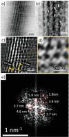
TEM imagery and Fourier analysis of the axial filaments isolated from spicules of the glass sponge M. chuni. b) Zoomed image of a) the TEM image that represents nanofibrillar organization of the selected area of isolated axial filament of M. chuni shows the cross‐linked nanoarchitecture (arrows) typical for F‐actin filaments, which form right‐handed, parallel, and staggered structures in all eukaryotes.[ 38 , 41 ] The FFT in (e) taken from (d) indicates different large periodicities typical for actin such as 5.9, 4.6, 3.7, 3.4, and 1.9 nm (see for details including statistical analysis Table S7, Supporting Information). Individual reflections are shown with red colored dotted lines. d) The axial filament lattice shows high similarity to that of actin standards reported by other authors.[ 37 , 40 ]
Figure 5
Figure 5
Latrunculin B‐mediated inhibition of siliceous spicules formation in S. lacustris demosponge. a) Light microscopy and SEM images of young sponge observed after hatching of gemmules under natural conditions shows the presence of c, arrows) glassy spicules. b) However, no spicules were observed in young sponges after the cultivation of the gemmules in the presence of latrunculin B (SEM image D), a well‐known inhibitor of actin polymerization in vivo (see also Figure S24, Supporting Information). TEM images of corresponding thin sections of sclerocytes confirm these observations and show f, arrow) the absence of spicules after treatment of gemmules with Latrunculin B in contrast to e) typical spicules formation under natural conditions (black, electron‐dense structures of biosilica, which surround the axial filaments of young spicules).

References

    1. Pisani D., Pett W., Dohrmann M., Feuda R., Rota‐Stabelli O., Philippe H., Lartillot N., Wörheide G., Proc. Natl. Acad. Sci. USA 2015, 112, 15402. - PMC - PubMed
    1. Telford M. J., Moroz L. L., Halanych K. M., Nature 2016, 529, 286. - PubMed
    1. Simion P., Philippe H., Baurain D., Jager M., Richter D. J., Di Franco A., Roure B., Satoh N., Quéinnec É., Ereskovsky A., Lapébie P., Corre E., Delsuc F., King N., Wörheide G., Manuel M., Curr. Biol. 2017, 27, 958. - PubMed
    1. Dohrmann M., Wörheide G., Sci. Rep. 2017, 7, 3599. - PMC - PubMed
    1. Systema Porifera: A Guide to the Classification of Sponges (Eds: Hooper J. N. A., van Soest R. W. M.), Kluwer Academic/Plenum Publishers, Dordrecht: 2002.

Publication types

LinkOut - more resources