. 2022 Feb 15;149(4):dev200035.
doi: 10.1242/dev.200035. Epub 2022 Feb 23.

Dlx1/2-dependent expression of Meis2 promotes neuronal fate determination in the mammalian striatum

Affiliations

Dlx1/2-dependent expression of Meis2 promotes neuronal fate determination in the mammalian striatum

Zihao Su et al. Development. .

Abstract

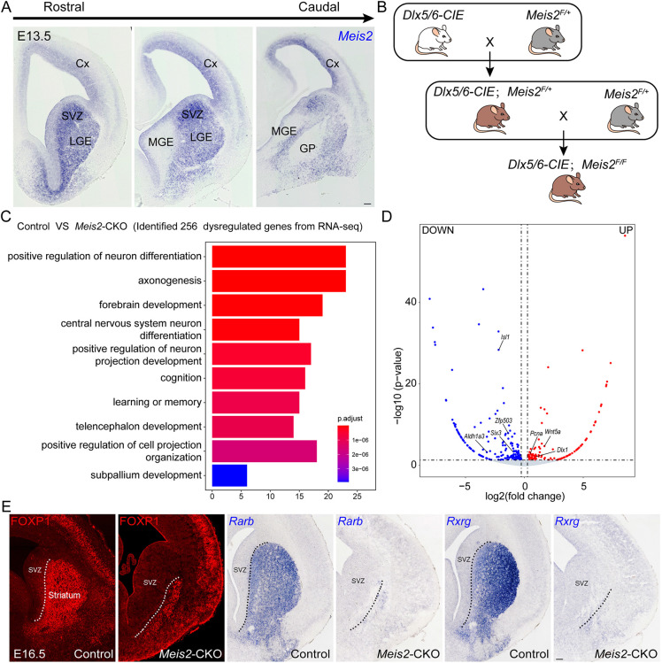
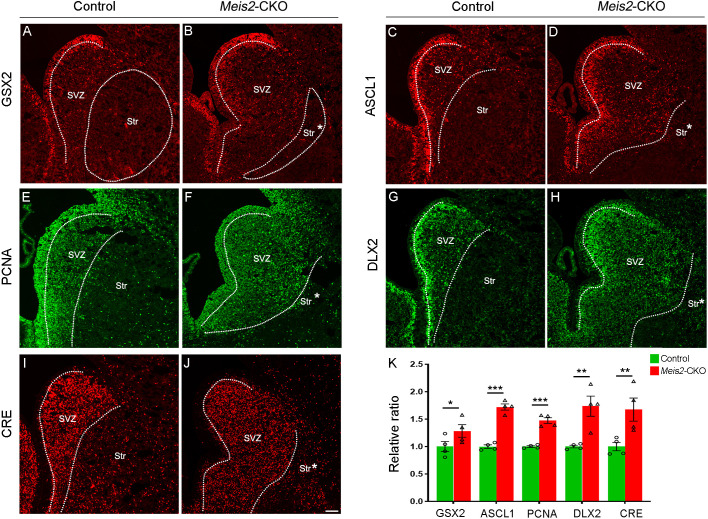
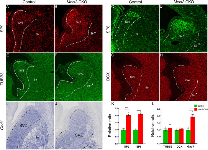
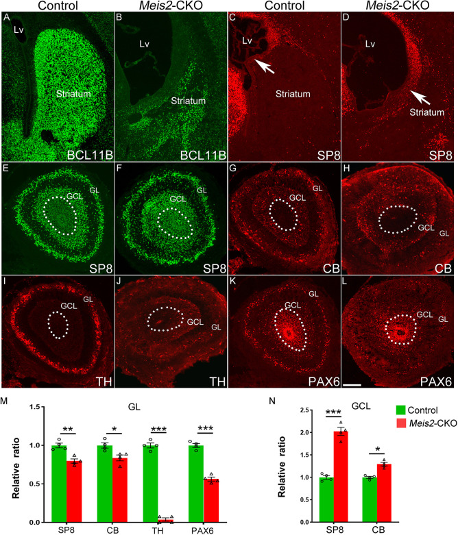
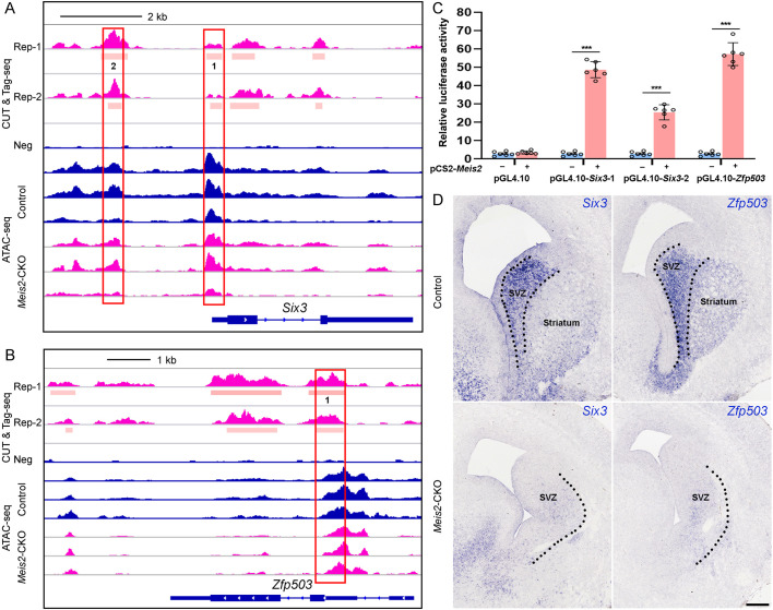
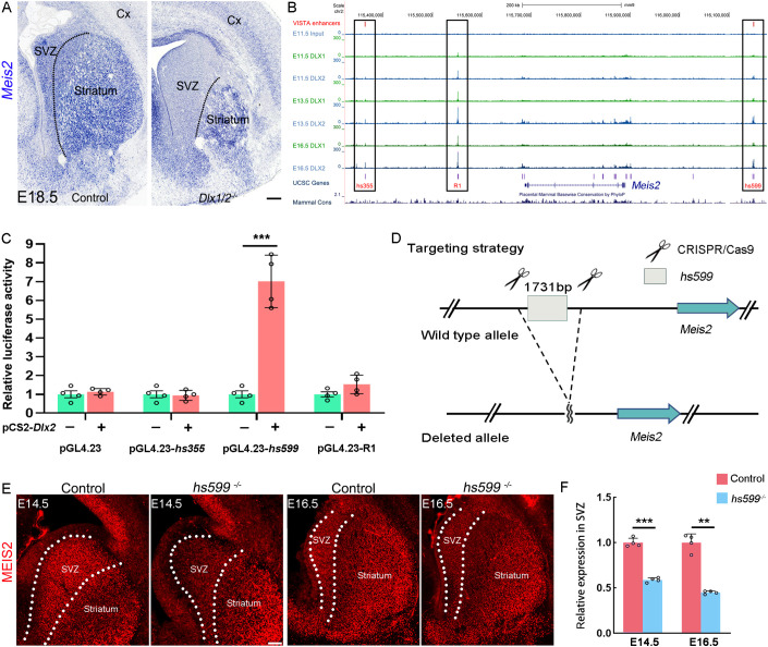
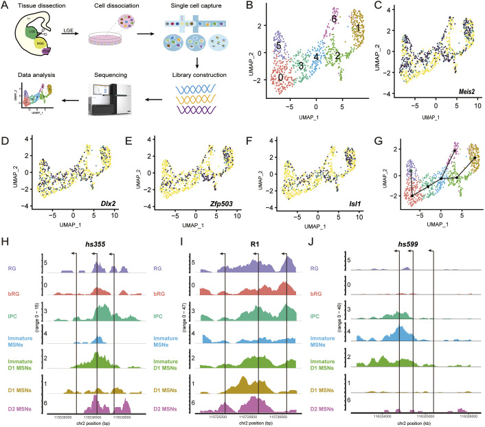
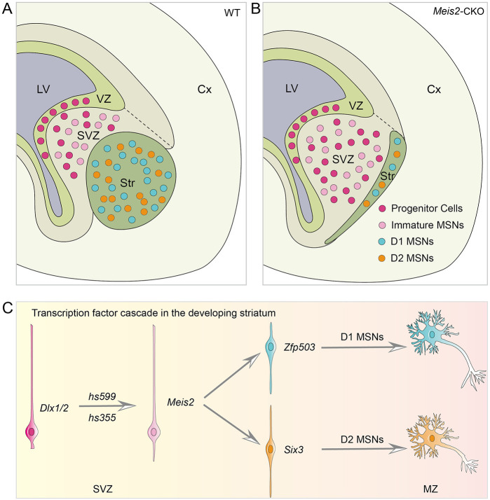
The striatum is a central regulator of behavior and motor function through the actions of D1 and D2 medium-sized spiny neurons (MSNs), which arise from a common lateral ganglionic eminence (LGE) progenitor. The molecular mechanisms of cell fate specification of these two neuronal subtypes are incompletely understood. Here, we found that deletion of murine Meis2, which is highly expressed in the LGE and derivatives, led to a large reduction in striatal MSNs due to a block in their differentiation. Meis2 directly binds to the Zfp503 and Six3 promoters and is required for their expression and specification of D1 and D2 MSNs, respectively. Finally, Meis2 expression is regulated by Dlx1/2 at least partially through the enhancer hs599 in the LGE subventricular zone. Overall, our findings define a pathway in the LGE whereby Dlx1/2 drives expression of Meis2, which subsequently promotes the fate determination of striatal D1 and D2 MSNs via Zfp503 and Six3.

Keywords: Dlx1/2; Meis2; Six3; Zfp503; LGE; Mouse; Striatum.


Conflict of interest statement

Competing interests The authors declare no competing or financial interests.

Figures

Fig. 1.
Fig. 1.
The majority of striatal MSNs are lost in Meis2-CKO mice. (A) Meis2 mRNA expression in a series of rostral-to-caudal sections at E13.5. Its expression is strongest in the LGE and septal progenitors and neurons (striatal primordium) and is remarkably low in the MGE and its derivatives. (B,C) Schematic showing crosses (B) and functional enrichment analysis (C) of DEGs in the LGE of Meis2-CKO versus control mice at E14.5. (D) Volcano plots showing the DEGs in the LGE at E14.5 between control and Meis2-CKO mice. (E) The expression of FOXP1, Rarb and Rxrg (three pan-neuronal markers of the MSNs) was significantly decreased in Meis2-CKO mice compared with control mice at E16.5. Dotted lines mark the border of the LGE SVZ and striatum. N=4 mice per group. Cx, cortex; GP, globus pallidus; LGE, lateral ganglionic eminence; MGE, medial ganglionic eminence; SVZ, subventricular zone. Scale bar: 100 µm for E.
Fig. 2.
Fig. 2.
Neural progenitors accumulate in the LGE SVZ of Meis2-CKO mice. (A-F) The number of cells expressing ventricular zone and SVZ markers, GSX2 (A,B), ASCL1 (C,D) and PCNA (E,F), were increased in a greatly expanded SVZ of Meis2-CKO mice at E16.5. Note that the mantle zone was smaller in Meis2-CKO mice than in control mice. (G-J) Likewise, the number of cells expressing SVZ and neuronal markers, DLX2 (G,H) and CRE (Dlx5/6) (I,J), were greatly increased in SVZ of the Meis2-CKO mice at E16.5. (K) The number of GSX2-, ASCL1-, PCNA-, DLX2- and CRE (Dlx5/6)-positive cells was significantly increased in Meis2-CKO mice compared with control mice. *P<0.05, **P<0.01, ***P<0.001 (one-way ANOVA followed by Tukey–Kramer post-hoc test). n=4 mice per group, mean±s.e.m. Dotted lines mark the border of the lateral ganglionic eminence SVZ and striatum. Str, striatum; Str*, presumptive striatum; SVZ, subventricular zone. Scale bar: 200 µm in J for A-J.
Fig. 3.
Fig. 3.
The abnormal differentiation MSNs accumulate in the LGE SVZ of Meis2-CKO mice. (A-D) The expression of the transcription factors SP8 (C,D) and SP9 (A,B) are increased in the LGE SVZ of the Meis2-CKO mice compared with the controls. (E-H) There are many immature neurons which express the pan-neuronal markers TUBB3 (E,F) and DCX (G,H) located in the LGE SVZ of Meis2-CKO and control mice. (I,J) The expression of Gad1 is increased in the LGE SVZ of the Meis2-CKO mice compared with the controls. (K,L) The quantification data of the above markers in the LGE SVZ. ***P<0.001 (one-way ANOVA followed by Tukey–Kramer post-hoc test). n=4 mice per group, mean±s.e.m. Dotted lines mark the border of the LGE SVZ and striatum. LGE, lateral ganglionic eminence; Str, striatum; Str*, presumptive striatum; SVZ, subventricular zone. Scale bar: 200 µm in J for A-J.
Fig. 4.
Fig. 4.
Meis2 is required for the development of olfactory bulb interneurons. (A,B) Compared with controls, the number of BCL11B-positive cells was reduced in Meis2-CKO mice at P0 in the striatum. (C,D) More SP8-positive cells were observed in the LGE SVZ of Meis2-CKO mice (arrows). (E-H) The cell number of the SP8- and CB-positive cells was increased in the GCL and decreased in the GL. (I,J) There was a significant reduction in TH-positive cells in the GL of the Meis2-CKO mice at P0. (K,L) The expression of PAX6 was significantly reduced in the GL. (M,N) Quantification of above experiments. *P<0.05, **P<0.01, ***P<0.001 (one-way ANOVA followed by Tukey–Kramer post-hoc test). n=4 mice per group, mean±s.e.m. Dotted lines mark the border of the olfactory bulb core. GCL, granule cell layer; GL, glomerular layer. Scale bar: 200 µm in L for A-L.
Fig. 5.
Fig. 5.
Meis2 is required for the differentiation of striatal MSNs. (A,B) Genomic regions of the Six3 (A) and Zfp503 (B) loci in the CUT&Tag-seq and ATAC-seq datasets. (C) MEIS2-activated transcription from the Six3 and Zfp503 promoters in a dual-luciferase assay of N2a cells. ***P<0.001 (unpaired two-tailed Student's t-test). n=6, mean±s.e.m. (D) In situ RNA hybridization analysis of Six3 and Zfp503 in control and Meis2-CKO mice at E16.5. Dotted lines mark the border of the lateral ganglionic eminence subventricular zone (SVZ) and striatum. Scale bar: 200 µm in D.
Fig. 6.
Fig. 6.
Dlx1/2 directly binds hs599 to promote Meis2 expression. (A) Meis2 expression was greatly reduced in the LGE SVZ of Dlx1/2 mutant mice. (B) DLX1/2 ChIP-seq showed that DLX1/2 directly bound to three distal regions near the Meis2 gene. (C) The dual-luciferase assay showed that DLX1/2 promoted the activity of hs599. (D) The CRISPR/Cas9 strategy was used to generate the hs599 mutant allele. (E,F) Meis2 expression was reduced in the LGE SVZ of the hs599 mutant mice at E14.5 and E16.5. N=4 mice per group. Dotted lines mark the border of the LGE SVZ and striatum. **P<0.01, ***P<0.001 (unpaired two-tailed Student's t-test). n=4, mean±s.e.m. Cx, cortex; LGE, lateral ganglionic eminence; SVZ, subventricular zone. Scale bar: 200 µm in A; 100 µm in E.
Fig. 7.
Fig. 7.
scATAC-seq revealed that hs599 exerted its function in a subpopulation of striatal MSNs. (A) Workflow of scATAC-seq of mouse LGE at E14.5. (B) Seven clusters (C0-C6) were identified and annotated according to marker gene accessibility. (C-F) UMAP plots show distinct chromatin accessibility profiles of marker genes. (G) The developmental trajectory construction scheme. (H-J) Genome tracks show normalized accessibility of three DLX1/2 binding regions with cluster-specific accessibility. Arrows represent the binding sites of DLX1/2. bRG, basal radial glial cells; Cx, cortex; IPC, intermediate progenitor cell; LGE, lateral ganglionic eminence; MGE, medial ganglionic eminence; MSN, medium-sized spiny neurons; POA, preoptic area; RG, radial glial cells.
Fig. 8.
Fig. 8.
A model of the mechanism by which Meis2 regulates striatal MSN development. (A,B) Proliferating and immature cells accumulated in the lateral ganglionic eminence SVZ in Meis2-CKO mice, compared with control mice. Notably, the Meis2-CKO mice have an enlarged SVZ and a small striatum. (C) The main transcription factor network regulating the fate determination of striatal MSNs. First, Dlx1/2 promote the expression of Meis2 by enhancers such as hs599 and hs355. Then, Meis2 promotes Zfp503 or Six3 expression to further determine precursor cell fate (D1 or D2 MSNs). Cx, cortex; LV, lateral ventricle; MSN, medium-sized spiny neurons; MZ, mantle zone; Str, striatum; SVZ, subventricular zone; VZ, ventricular zone.

Similar articles

Cited by

References

    1. Anderson, S. A., Qiu, M., Bulfone, A., Eisenstat, D. D., Meneses, J., Pedersen, R. and Rubenstein, J. L. R. (1997). Mutations of the homeobox genes Dlx-1 and Dlx-2 disrupt the striatal subventricular zone and differentiation of late born striatal neurons. Neuron 19, 27-37. 10.1016/S0896-6273(00)80345-1 - DOI - PubMed
    1. Arlotta, P., Molyneaux, B. J., Jabaudon, D., Yoshida, Y. and Macklis, J. D. (2008). Ctip2 controls the differentiation of medium spiny neurons and the establishment of the cellular architecture of the striatum. J. Neurosci. 28, 622-632. 10.1523/JNEUROSCI.2986-07.2008 - DOI - PMC - PubMed
    1. Banan, M. (2020). Recent advances in CRISPR/Cas9-mediated knock-ins in mammalian cells. J. Biotechnol. 308, 1-9. 10.1016/j.jbiotec.2019.11.010 - DOI - PubMed
    1. Berenguer, M. and Duester, G. (2021). Role of retinoic acid signaling, FGF signaling and Meis genes in control of limb development. Biomolecules 11, 80. 10.3390/biom11010080 - DOI - PMC - PubMed
    1. Bocchi, V. D., Conforti, P., Vezzoli, E., Besusso, D., Cappadona, C., Lischetti, T., Galimberti, M., Ranzani, V., Bonnal, R. J. P., De Simone, M.et al. (2021). The coding and long noncoding single-cell atlas of the developing human fetal striatum. Science 372, eabf5759. 10.1126/science.abf5759 - DOI - PubMed

Publication types

MeSH terms