. 2022 Mar 31;42(3):BSR20212457.
doi: 10.1042/BSR20212457.

Insights into pathological mechanisms and interventions revealed by analyzing a mathematical model for cone metabolism

Affiliations

Insights into pathological mechanisms and interventions revealed by analyzing a mathematical model for cone metabolism

Atanaska Dobreva et al. Biosci Rep. .

Abstract

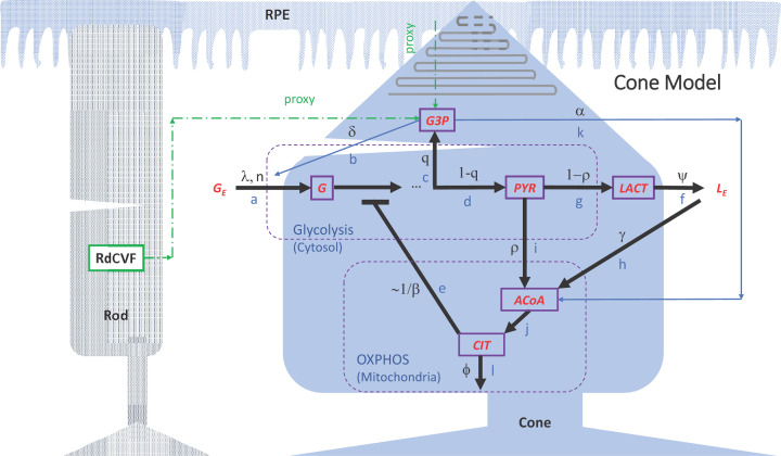
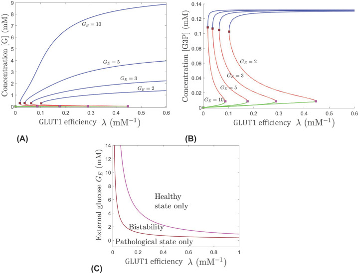
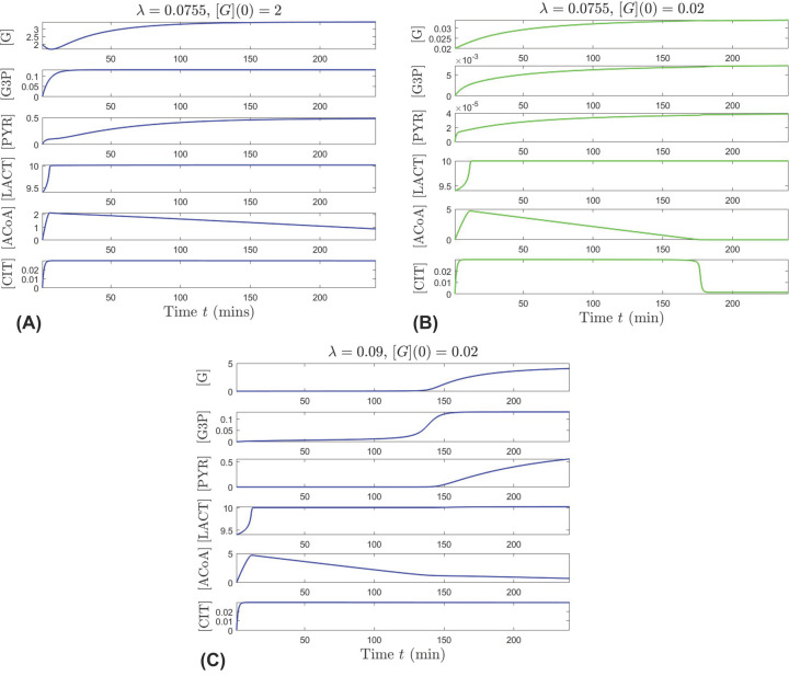
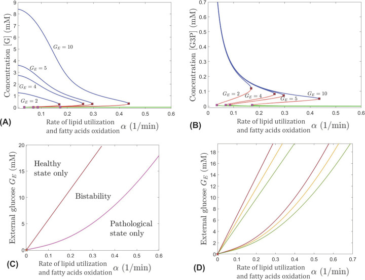
This work analyzes a mathematical model for the metabolic dynamics of a cone photoreceptor, which is the first model to account for energy generation from fatty acids oxidation of shed photoreceptor outer segments (POS). Multiple parameter bifurcation analysis shows that joint variations in external glucose, the efficiency of glucose transporter 1 (GLUT1), lipid utilization for POS renewal, and oxidation of fatty acids affect the cone's metabolic vitality and its capability to adapt under glucose-deficient conditions. The analysis further reveals that when glucose is scarce, cone viability cannot be sustained by only fueling energy production in the mitochondria, but it also requires supporting anabolic processes to create lipids necessary for cell maintenance and repair. In silico experiments are used to investigate how the duration of glucose deprivation impacts the cell without and with a potential GLUT1 or oxidation of fatty acids intervention as well as a dual intervention. The results show that for prolonged duration of glucose deprivation, the cone metabolic system does not recover with higher oxidation of fatty acids and requires greater effectiveness of GLUT1 to recover. Finally, time-varying global sensitivity analysis (GSA) is applied to assess the sensitivity of the model outputs of interest to changes and uncertainty in the parameters at specific times. The results reveal a critical temporal window where there would be more flexibility for interventions to rescue a cone cell from the detrimental consequences of glucose shortage.

Keywords: Bifurcation Analysis; Metabolism; Oxidation of fatty acids; Photoreceptors; Retina; Sensitivity Analysis.


Conflict of interest statement

The authors declare that there are no competing interests associated with the manuscript.

Figures

Figure 1
Figure 1. Model diagram of key metabolic pathways within a single cone photoreceptor
The diagram was modified from Camacho et al. (2020) [25] to show interactions of the cone with the RPE and a rod photoreceptor (dashed green arrows). Parameters α and δ used in the cone model are defined as proxies for interactions with the RPE and rods. To isolate the impact on the cone cell of changes in glucose modulation by the RPE in response to reduction in RPE’s metabolic fuel, the pathways for energy generation in the RPE and rods via β-HB are not included. Model variables are represented by boxes with purple border: glucose (G), G3P, PYR, lactate (LACT), ACoA, and CIT. Metabolic pathways are labeled with blue letters: (a,b) Glucose transport and uptake (without and with RdCVF); (c) G3P synthesis; (d) Pyruvate synthesis; (e) Glycolysis inhibition; (f) Outward lactate transport; (g) Lactate synthesis; (h) Inward lactate transport for ACoA production; (i) Pyruvate conversion into ACoA; (j) Citrate synthesis; (k) ACoA synthesis from β-HB; (l) Diversion of citrate to the cytosol and other pathways. Pathway-associated parameters are labeled with black letters, and external glucose and lactate are denoted by GE and LE, respectively. Parameter descriptions are provided in Table A1 in Appendix A.
Figure 2
Figure 2. Bifurcations in dynamics with respect to the glucose transport factor λ, which serves as a quantifier for the efficiency of GLUT1, for different levels of the external RPE-mediated glucose GE
Evolution of the [G] (A) and [G3P] (B) components of the system’s equilibria, as λ is increased; blue (healthy) and green (pathological) curves are stable while red is unstable. Different curves represent the equilibrium curves for different levels of GE, from right to left: GE = 2, 3, 5, and 10 mM. Two saddle node transitions occur along each equilibrium curve for all values of GE. (C) Regions of unistability and bistability delimited in the (λ, GE) slice of the parameter space. The value of α was fixed to 0.2 min−1.
Figure 3
Figure 3. Transition out of the bistability regime, as the glucose transport factor λ that quantifies the efficiency of GLUT1 increases (top panels for λ = 0.0755 vs. bottom panel for λ = 0.09) under high external glucose level GE = 11.5 mM
Each panel shows the simultaneous evolution of all six components for one solution of the system, for initial conditions fixed at [G3P](0) = 0, [PYR](0) = 0, [LACT](0) = 9.4, [ACoA](0) = 0, [CIT](0) = 0 and parameters (other than λ) set to their nominal values given in Table 1. Blue represents solutions which converge to the healthy state, and green represents solutions that converge to the pathological state. For λ = 0.0755 mM−1, the system is in the bistability window; it converges to the healthy state when starting from higher internal glucose [G](0) = 2 mM (A) and to the pathological state when starting from low internal glucose [G](0) = 0.02 mM (B). For λ = 0.09 mM−1, the system has a unique healthy equilibrium, and converges to it even when starting from very low internal glucose [G](0) = 0.02 mM (C).
Figure 4
Figure 4. Bifurcations in dynamics with respect to α, for different levels of external RPE-mediated glucose GE
Top panels: Evolution of the [G] (A) and [G3P] (B) components of the system’s equilibria, as α is increased; blue (healthy) and green (pathological) curves are stable while red is unstable. Different curves represent the equilibrium curves for different levels of GE, from left to right: GE = 2, 4, 5, and 10 mM. Two saddle node bifurcations occur along each equilibrium curve for all values of GE. (C) Regions of unistability and bistability delimited (as labeled) in the (α, GE) parameter slice by two saddle node curves, shown in brown and in pink, meeting at a cusp point near the origin, marked with a star. The value of λ is set to 0.0755 mM−1. (D) Delimitation between healthy/bistability/pathological regimes in the (α, GE) parameter plane for three different values of the glucose transport factor: λ = 0.065 mM−1 (red curve), λ = 0.075 mM−1 (orange curve), and λ = 0.09 mM−1 (green curve).
Figure 5
Figure 5. In glucose deprivation conditions, supplementation of exogenous fuels from fatty acids oxidation and lactate conversion into pyruvate prompts short-term rescue of oxidative metabolism (as reflected by the purple curves for CIT and ACoA)
For all curves in panels (A,B), GE = [G] throughout the simulation, eliminating the glucose gradient differential and thus blocking glucose uptake. Additionally, [G](0) = 0.5, [LACT](0) = 9.4 and different initial concentrations of G3P and external lactate, LE, are considered; [G3P](0) = 0 and LE = 9.4 (orange curves in both panels), [G3P](0) = 2 and LE = 9.4 (purple curves in panel A), and [G3P](0) = 0 and LE = 9.92 (purple curves in panel B). All other initial conditions are set to 0, and all other parameters are kept at the nominal values presented in Table 1.
Figure 6
Figure 6. Impact of glucose shutdown duration and interventions
(A,B) Effect of temporarily shutting down glucose uptake. (A) Time evolution of the six metabolites when α = 0.1 and λ = 0.0755, under three different circumstances: GE = 10 mM (blue curves); GE = [G] (green curves); GE = [G] only between t = 40 min and t = 130 min, and GE = 10 otherwise (red curves). (B) Similar trajectories to the ones illustrated in (A), for a higher α = 0.18. (C,D) Strategies for restoring healthy metabolite levels after temporarily shutting down glucose uptake. (C) Restore GE earlier. Time evolution of the six metabolites when α = 0.18 and λ = 0.0755, when GE = [G] between t = 40 min and t = 130 min (green curve), and when shutdown ends early at t = 100 min (red curve). (D) Increase λ. Time evolution of the six metabolites when α = 0.18 and λ = 0.09, under the same three conditions as in panels (A,B): GE = 10 mM (blue curves); GE = [G] (green curves); GE = [G] only between t = 40 min and t = 130 min, and GE = 10 otherwise (red curves). For all panels, the trigger times are shown as dots along the corresponding curves; LE = 9.4 mM and the other parameters are kept at their nominal values in Table 1; the initial conditions are [G](0) = 5 mM, [LACT](0) = 9.4 mM, and the other components set to zero.
Figure 7
Figure 7. Time-dependent GSA results for low initial glucose level
Left panels present time-dependent results of PRCC and right panels present time-dependent results of eFAST, both for initial glucose concentration of [G](0) = 0.02 mM. Two outcomes of interest are considered, presented in rows 1,2: intracellular concentrations of glucose ([G]) and G3P ([G3P]). The gray bands on the left panels indicate PRCC values which are not statistically significant (P-value ≥0.001).
Figure 8
Figure 8. Time-dependent GSA results for higher initial glucose level
Left panels present time-dependent results of PRCC and right panels present time-dependent results of eFAST, both for initial glucose concentration of [G](0) = 2 mM. Two outcomes of interest are considered, presented in rows 1,2: intracellular concentrations of glucose ([G]) and G3P ([G3P]). The gray bands on the left panels indicate PRCC values which are not statistically significant (P-value ≥0.001).

References

    1. Cheng S.-Y., Cipi J., Ma S., Hafler B.P., Kanadia R.N., Brush R.S.et al. . (2020) Altered photoreceptor metabolism in mouse causes late stage age-related macular degeneration like pathologies. Proc. Natl. Acad. Sci. U.S.A. 117, 13094–13104 10.1073/pnas.2000339117 - DOI - PMC - PubMed
    1. Camacho E.T., Brager D., Elachouri G., Korneyeva T., Millet-Puel G., Sahel J.-A.et al. . (2019) A mathematical analysis of aerobic glycolysis triggered by glucose uptake in cones. Sci. Rep. 9, 4162 10.1038/s41598-019-39901-z - DOI - PMC - PubMed
    1. Leveillard T. and Sahel J.-A. (2017) Metabolic and redox signaling in the retina. Cell. Mol. Life Sci. 74, 3649–3665 10.1007/s00018-016-2318-7 - DOI - PMC - PubMed
    1. Chertov A.O., Holzhausen L., Kuok I.T., Couron D., Parker E.D., Linton J.D.et al. . (2011) Roles of glucose in photoreceptor survival. J. Biol. Chem. 286, 34700–34711 10.1074/jbc.M111.279752 - DOI - PMC - PubMed
    1. Camacho E.T., Leveillard T., Sahel J.-A. and Wirkus S. (2016) Mathematical model of the role of rdcvf in the coexistence of rods and cones in a healthy eye. Bull. Math. Biol. 78, 1394–1409 10.1007/s11538-016-0185-x - DOI - PubMed

Publication types

MeSH terms