. 2022 May;28(5):749-760.
doi: 10.1111/cns.13807. Epub 2022 Feb 14.

Inhibition of phosphodiesterase-4 in the spinal dorsal horn ameliorates neuropathic pain via cAMP-cytokine-Cx43 signaling in mice

Affiliations

Inhibition of phosphodiesterase-4 in the spinal dorsal horn ameliorates neuropathic pain via cAMP-cytokine-Cx43 signaling in mice

Fang-Fang Zhang et al. CNS Neurosci Ther. 2022 May.

Abstract

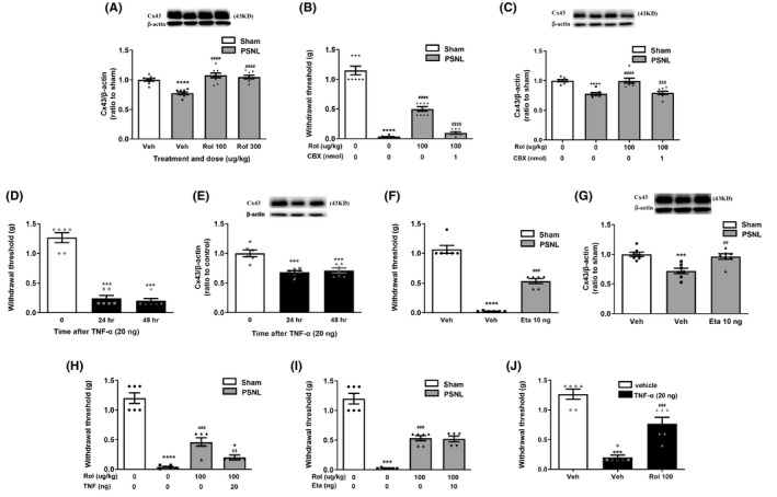
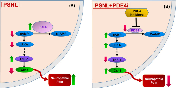
Background: The spinal phosphodiesterase-4 (PDE4) plays an important role in chronic pain. Inhibition of PDE4, an enzyme catalyzing the hydrolysis of cyclic adenosine monophosphate AMP (cAMP), produces potent antinociceptive activity. However, the antinociceptive mechanism remains largely unknown. Connexin43 (Cx43), a gap junction protein, has been shown to be involved in controlling pain transduction at the spinal level; restoration of Cx43 expression in spinal astrocytes to the normal levels reduces nerve injury-induced pain. Here, we evaluate the novel mechanisms involving spinal cAMP-Cx43 signaling by which PDE4 inhibitors produce antinociceptive activity.

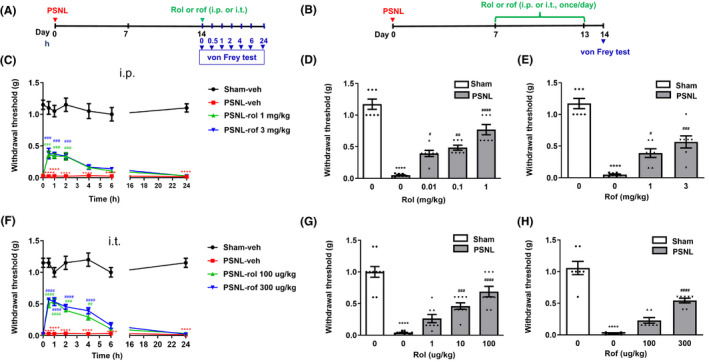
Methods: First, we determined the effect of PDE4 inhibitors rolipram and roflumilast on partial sciatic nerve ligation (PSNL)-induced mechanical hypersensitivity. Next, we observed the role of cAMP-Cx43 signaling in the effect of PDE4 inhibitors on PSNL-induced mechanical hypersensitivity.

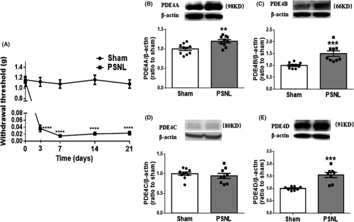
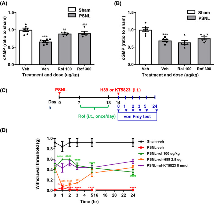
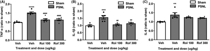
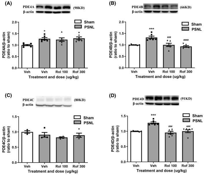
Results: Single or repeated, intraperitoneal or intrathecal administration of rolipram or roflumilast significantly reduced mechanical hypersensitivity in mice following PSNL. In addition, repeated intrathecal treatment with either of PDE4 inhibitors reduced PSNL-induced downregulation of cAMP and Cx43, and upregulation of proinflammatory cytokines tumor necrosis factor-α (TNF-α) and interleukin-1β. Furthermore, the antinociceptive effects of PDE4 inhibitors were attenuated by the protein kinase A (PKA) inhibitor H89, TNF-α, or Cx43 antagonist carbenoxolone. Finally, PSNL-induced upregulation of PDE4B and PDE4D, especially the PDE4B subtype, was reduced by treatment with either of the PDE4 inhibitors.

Conclusions: The results suggest that the antinociceptive effect of PDE4 inhibitors is contributed by increasing Cx43 expression via cAMP-PKA-cytokine signaling in the spinal dorsal horn.

Keywords: connexin43; neuropathic pain; phosphodiesterase-4; roflumilast; rolipram.


Conflict of interest statement

The authors declare that they have no conflicts of interest.

Figures

FIGURE 1
FIGURE 1
Partial sciatic nerve ligation (PSNL) produced mechanical hypersensitivity and increased expression of PDE4 subtypes in the spinal dorsal horn in mice. (A) Ipsilateral hind paw withdrawal thresholds (g) over time measured with von Frey filaments. Mice were tested before (0 day), and 3, 7, 14, and 21 days after PSNL or sham surgery. (B‐E) The expression of PDE4A‐D in the ipsilateral spinal dorsal horn of mice subjected to PSNL or sham, quantified by Western blotting analysis 14 days after surgery. The optic densities of PDE4A‐D were normalized to those of sham controls. Data shown are means ± SEM. ** p < 0.01, *** p < 0.001, **** p < 0.0001 vs. corresponding sham; n = 10 (A) or 8–10 per group (B‐E)
FIGURE 2
FIGURE 2
Intraperitoneal or intrathecal treatment with PDE4 inhibitors ameliorated mechanical hypersensitivity induced by PSNL in mice. (A) Schedule of acute PDE4 inhibitor administration and testing. Rolipram (rol), roflumilast (rof), or vehicle (veh) was intraperitoneal (i.p.) or intrathecally (i.t.) injected 14 days after PSNL; hind paw withdrawal thresholds (g) were determined using the von Frey test at 0, 0.5, 1, 2, 4, 6, and 24 h after the injection. (B) Schedule of repeated injections of PDE4 inhibitors. Rolipram, roflumilast, or vehicle was injected (i.p. or i.t.) 7 times (Days 7, 8, 9, 10, 11, 12, and 13 after PSNL surgery). Mice were tested for withdrawal thresholds using the von Frey test on Day 14. (C) Changes in hind paw withdrawal thresholds overtime after acute treatment (i.p.) with rolipram (1 mg/kg), roflumilast (3 mg/kg), or vehicle. (D, E) Effects of repeated treatment (i.p.) with rolipram (0.01, 0.1, or 1 mg/kg) (D) or roflumilast (1 or 3 mg/kg) (E) on hind paw withdrawal thresholds (g) 14 days following PSNL or sham surgery. (F) Changes in withdrawal thresholds (g) following single, intrathecal injections of rolipram (100 µg/kg), roflumilast (300 µg/kg), or vehicle at 0, 0.5, 1, 2, 4, 6, and 24 h after the injection. (G, H) Effects of repeated intrathecal treatment with rolipram (1, 10, or 100 µg/kg) (G) or roflumilast (100 or 300 µg/kg) (H) on hind paw withdrawal thresholds (g) measured by the von Frey test 14 days following PSNL surgery. Data shown are means ± SEM. **** p < 0.0001 vs. sham with vehicle; # p < 0.05, ## < 0.01, ### p < 0.001, #### p < 0.0001 vs. PSNL with vehicle; n = 7–10 per group
FIGURE 3
FIGURE 3
The role of rolipram or roflumilast on cAMP‐PKA signaling in the spinal dorsal horn. (A) Effect of repeated intrathecal treatment with vehicle (veh), rolipram (rol, 100 µg/kg), or roflumilast (rof, 300 µg/kg) on cAMP (A) and cGMP (B) levels in the spinal dorsal horn of PSNL mice. Rolipram (100 µg/kg), roflumilast (300 µg/kg), or vehicle was intrathecally injected once a day for 7 days following the schedule described in Figure 2B. Mice were sacrificed, and the spinal dorsal horn was collected 14 days after PSNL surgery for the determination of cAMP and cGMP levels using ELISA kits. (C) Schedule of drug injections and testing. Rolipram or vehicle was intrathecally injected once a day for 7 days (i.e., 7–13 days after PSNL); H89 or KT5823 was intrathecally injected on Day 14. The hind paw withdrawal thresholds were determined using the von Frey test at 0, 1, 2, 3, 5, and 24 h after the injection. (D) Effects of rolipram (100 µg/kg) on hind paw withdrawal thresholds (g) over time with or without the intrathecal injection of H89 (2.5 µg/5 µl) or KT5823 (8 nmol) in PSNL mice. Data shown are means ± SEM. ** p < 0.01, *** p < 0.001, **** p < 0.0001 vs. sham with vehicle; ## p < 0.01, ### p < 0.001, #### p < 0.0001 vs. PSNL with vehicle; $$$ p < 0.001, $$$$ p < 0.0001 vs. PSNL with rolipram; n = 5–6 per group
FIGURE 4
FIGURE 4
Effects of rolipram or roflumilast on levels of TNF‐α (A), IL‐1β (B), and IL‐6 (C) in the spinal dorsal horn of PSNL mice. Rolipram (rol, 100 µg/kg), roflumilast (rof, 300 µg/kg), or vehicle (veh) was intrathecally injected once a day for 7 days following the schedule described in Figure 2B. Mice were sacrificed, and the spinal dorsal horn was collected 14 days after PSNL surgery for determination of TNF‐α (A), IL‐1β (B), and IL‐6 (C) levels using ELISA kits. Data shown are means ± SEM. ** p < 0.01, *** p < 0.001, **** p < 0.0001 vs. sham with vehicle; ## p < 0.01, #### p < 0.0001 vs. PSNL with vehicle; n = 5–6 per group
FIGURE 5
FIGURE 5
Effects of PDE4 inhibitors on cytokine‐Cx43 signaling in the spinal dorsal horn of PSNL mice. (A) Expression of Cx43 by Western blotting in the spinal dorsal horn of PSNL mice treated with vehicle (veh), rolipram (rol, 100 µg/kg), or roflumilast (rof, 300 µg/kg). Rolipram, roflumilast, or vehicle was intrathecally injected once a day for 7 days, as identified in Figure 2B. (B, C) Rolipram (rol, 100 µg/kg) was intrathecal injected 7 times (7, 8, 9, 10, 11, 12, and 13 days after PSNL surgery) and CBX (1 nmol) was intrathecally injected once on Day 13. Hind paw withdrawal thresholds (g) (B) and Cx43 expression in the spinal dorsal horn (C) were measured using the von Frey test and western blot 14 days following PSNL surgery. (D, E) TNF‐α decreased hind paw withdrawal thresholds (D) and Cx43 expression in the spinal dorsal horn (E) assessed 24 and 48 h after the last intrathecal injection. TNF‐α (20 ng) was intrathecally injected, and hind paw withdrawal thresholds were measured 24 and 48 h after the injection (D and E). (F, G) The effect of etanercept on hind paw withdrawal thresholds (F) and Cx43 expression (G) in PSNL mice. Etanercept (Eta, 10 ng) was intrathecally injected 7 times (7, 8, 9, 10, 11, 12, and 13 days after PSNL surgery), and hind paw withdrawal thresholds were measured on Day 14 (F and G). The spinal dorsal horn was collected for Western blotting for Cx43 expression immediately after the behavioral test. (H) TNF‐α blocked the antinociceptive effect of rolipram. Rolipram (rol, 100 µg/kg) was intrathecally injected 7 times (7, 8, 9, 10, 11, 12, and 13 days after PSNL surgery), and TNF‐α was singly and intrathecally injected on the 13th day. (I) Etanercept did not affect the antinociceptive effect of rolipram. Rolipram (100 µg/kg) was co‐injected intrathecally with etanercept (Eta, 10 ng) 7 times (7, 8, 9, 10, 11, 12, and 13 days after PSNL surgery). For both (H) and (I), mice were tested for hind paw withdrawal thresholds (g) 14 days post‐PSNL after the last intrathecal injections of rolipram with TNF‐α (20 ng) or Eta using the von‐Frey test. (J) Rolipram blocked the nociceptive effect of TNF‐α. Rolipram (100 µg/kg/day) was intrathecally injected for 5 days and TNF‐α (20 ng) injected 2 days before the hind paw withdrawal thresholds were measured using the von Frey test. Data shown are means ± SEM. ** p < 0.01, *** p < 0.001, **** p < 0.0001, vs. vehicle or sham with vehicle; ## p < 0.01, ### p < 0.001, ### p < 0.001 vs. PSNL with vehicle; n = 3 (C) or 9 per group (A). $$$ p < 0.001, $$$$ p < 0.0001 vs. PSNL with rolipram. n = 5–9 per group
FIGURE 6
FIGURE 6
Effects of PDE4 inhibitors on expression of PDE4 subtypes in the spinal dorsal horn in PSNL mice. (A–D) Effect of repeated intrathecal injections of rolipram or roflumilast on expression of PDE4A (A), PDE4B (B), PDE4C (C), and PDE4D (D) in the spinal dorsal horn using Western blotting. Rolipram (rol, 100 µg/kg) or roflumilast (rof, 300 µg/kg) was given following the schedule in Figure 2B. Data shown are means ± SEM. * p < 0.05, *** p < 0.001, vs. sham with vehicle; ### p < 0.001, #### p < 0.0001 vs. PSNL with vehicle; n = 6 per group
FIGURE 7
FIGURE 7
Schematic of the mechanisms by which PDE4 inhibitors attenuate neuropathic pain in PSNL mice. (A) In the spinal dorsal horn in PSNL mice, expression of PDE4 is induced, which causes decreases in cAMP levels and PKA activity, leading to activation of proinflammatory cytokines such as TNF‐α and downregulation of Cx43, eventually resulting in neuropathic pain. (B) Following treatment with PDE4 inhibitors (PDE4i) in PSNL mice, cAMP‐PKA signaling is activated and cytokines such as TNF‐α are suppressed, leading to increases in Cx43 and eventual suppression of neuropathic pain. PSNL, partial sciatic nerve ligation; PDE4, phosphodiesterase‐4; 5′‐AMP, 5′‐adenosine monophosphate; cAMP, cyclic AMP; PKA, protein kinase A; TNF‐α, tumor necrosis factor‐α; Cx43, connexin43

Similar articles

Cited by

References

    1. Colloca L, Ludman T, Bouhassira D, et al. Neuropathic pain. Nat Rev Dis Primers. 2017;3:17002. - PMC - PubMed
    1. Grace PM, Hutchinson MR, Maier SF, Watkins LR. Pathological pain and the neuroimmune interface. Nat Rev Immunol. 2014;14:217‐231. - PMC - PubMed
    1. Kobayashi Y, Kiguchi N, Maeda T, Ozaki M, Kishioka S. The critical role of spinal ceramide in the development of partial sciatic nerve ligation‐induced neuropathic pain in mice. Biochem Biophys Res Commun. 2012;421:318‐322. - PubMed
    1. Même W, Calvo CF, Froger N, et al. Proinflammatory cytokines released from microglia inhibit gap junctions in astrocytes: potentiation by beta‐amyloid. FASEB J. 2006;20:494‐496. - PubMed
    1. Zhang FF, Morioka N, Nakashima‐Hisaoka K, Nakata Y. Spinal astrocytes stimulated by tumor necrosis factor‐ α and/or interferon‐γ attenuate connexin 43‐gap junction via c‐jun terminal kinase activity. J Neurosci Res. 2013;91:745‐756. - PubMed

MeSH terms