

Sustainable low-field cardiovascular magnetic resonance in changing healthcare systems
- PMID: 35157038
- PMCID: PMC9159744
- DOI: 10.1093/ehjci/jeab286
Sustainable low-field cardiovascular magnetic resonance in changing healthcare systems
Abstract
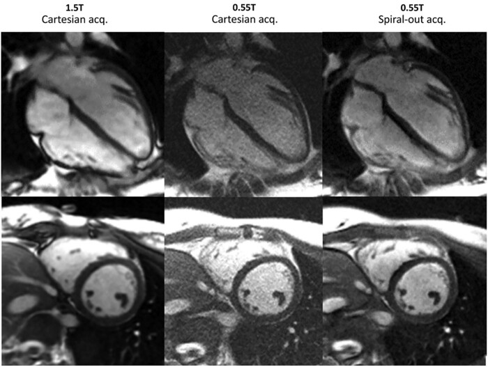
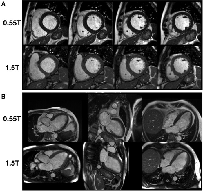
Cardiovascular disease continues to be a major burden facing healthcare systems worldwide. In the developed world, cardiovascular magnetic resonance (CMR) is a well-established non-invasive imaging modality in the diagnosis of cardiovascular disease. However, there is significant global inequality in availability and access to CMR due to its high cost, technical demands as well as existing disparities in healthcare and technical infrastructures across high-income and low-income countries. Recent renewed interest in low-field CMR has been spurred by the clinical need to provide sustainable imaging technology capable of yielding diagnosticquality images whilst also being tailored to the local populations and healthcare ecosystems. This review aims to evaluate the technical, practical and cost considerations of low field CMR whilst also exploring the key barriers to implementing sustainable MRI in both the developing and developed world.
Keywords: Global Health; Low field; MRI; Sustainable; Technology.
© The Author(s) 2022. Published by Oxford University Press on behalf of the European Society of Cardiology.
Figures
Similar articles
-
A comparison of cardiovascular imaging practices in Africa, North America, and Europe: two faces of the same coin.Eur Heart J Imaging Methods Pract. 2023 Jul 10;1(1):qyad005. doi: 10.1093/ehjimp/qyad005. eCollection 2023 May. Eur Heart J Imaging Methods Pract. 2023. PMID: 39044787 Free PMC article.
-
Noninvasive rapid cardiac magnetic resonance for the assessment of cardiomyopathies in low-middle income countries.Expert Rev Cardiovasc Ther. 2021 May;19(5):387-398. doi: 10.1080/14779072.2021.1915130. Epub 2021 May 24. Expert Rev Cardiovasc Ther. 2021. PMID: 33836619 Review.
-
Challenges and opportunities for early career medical professionals in cardiovascular magnetic resonance (CMR) imaging: a white paper from the Society for Cardiovascular Magnetic Resonance.J Cardiovasc Magn Reson. 2023 Nov 16;25(1):65. doi: 10.1186/s12968-023-00968-3. J Cardiovasc Magn Reson. 2023. PMID: 37968709 Free PMC article. Review.
-
Improving cardiovascular magnetic resonance access in low- and middle-income countries for cardiomyopathy assessment: rapid cardiovascular magnetic resonance.Eur Heart J. 2022 Jul 7;43(26):2496-2507. doi: 10.1093/eurheartj/ehac035. Eur Heart J. 2022. PMID: 35139531 Free PMC article.
-
Cardiovascular magnetic resonance for the evaluation of patients with cardiovascular disease: An overview of current indications, limitations, and procedures.Hellenic J Cardiol. 2023 Mar-Apr;70:53-64. doi: 10.1016/j.hjc.2023.01.003. Epub 2023 Jan 24. Hellenic J Cardiol. 2023. PMID: 36706867 Review.
Cited by
-
Feasibility of strain-encoded magnetic resonance at 0.55T.J Cardiovasc Magn Reson. 2025 Summer;27(1):101870. doi: 10.1016/j.jocmr.2025.101870. Epub 2025 Feb 25. J Cardiovasc Magn Reson. 2025. PMID: 40015457 Free PMC article.
-
Low-field MRI: A report on the 2022 ISMRM workshop.Magn Reson Med. 2023 Oct;90(4):1682-1694. doi: 10.1002/mrm.29743. Epub 2023 Jun 22. Magn Reson Med. 2023. PMID: 37345725 Free PMC article. Review.
-
Highly efficient image navigator based 3D whole-heart cardiac MRA at 0.55T.Magn Reson Med. 2025 Feb;93(2):689-698. doi: 10.1002/mrm.30316. Epub 2024 Oct 16. Magn Reson Med. 2025. PMID: 39415543 Free PMC article.
-
Observations of Cardiovascular Disease in Patients With Challenges to Health Care Access: A CMR Study.JACC Adv. 2024 Jul 13;3(8):101084. doi: 10.1016/j.jacadv.2024.101084. eCollection 2024 Aug. JACC Adv. 2024. PMID: 39105115 Free PMC article. No abstract available.
-
The environmental effects of non-invasive cardiac imaging.Am Heart J Plus. 2024 Sep 24;46:100463. doi: 10.1016/j.ahjo.2024.100463. eCollection 2024 Oct. Am Heart J Plus. 2024. PMID: 39399577 Free PMC article.
References
-
- Organization WH. Cardiovascular Diseases (CVDs)2017. https://www.who.int/en/news-room/fact-sheets/detail/cardiovascular-disea... (1 June 2021, date last accessed).
-
- Rosengren A, Smyth A, Rangarajan S, Ramasundarahettige C, Bangdiwala SI, AlHabib KF. et al. Socioeconomic status and risk of cardiovascular disease in 20 low-income, middle-income, and high-income countries: the Prospective Urban Rural Epidemiologic (PURE) study. Lancet Glob Health 2019;7:e748–60. - PubMed
-
- Celermajer DS, Chow CK, Marijon E, Anstey NM, Woo KS.. Cardiovascular disease in the developing world: prevalences, patterns, and the potential of early disease detection. J Am Coll Cardiol 2012;60:1207–16. - PubMed
-
- United Nations. Transforming Our World: The 2030 Agenda for Sustainable Development. 2015. https://sustainabledevelopment.un.org/content/documents/21252030%20Agend... (1 June 2021, date last accessed).
