Second and third trimester estimation of gestational age using ultrasound or maternal symphysis-fundal height measurements: A systematic review
- PMID: 35157348
- PMCID: PMC9545821
- DOI: 10.1111/1471-0528.17123
Second and third trimester estimation of gestational age using ultrasound or maternal symphysis-fundal height measurements: A systematic review
Abstract
Many vulnerable women seek antenatal care late in pregnancy. How should gestational age be determined? We examine all available studies estimating GA >20 weeks. Ultrasound is much better than fundal height, and using cerebellar measurement appears to be the most accurate.
Linked article: This article is commented on by Philip J. Steer, pp. 1459 in this issue. To view this minicommentary visit
Background: Accurate assessment of gestational age (GA) is important for managing pregnancies at an individual level and monitoring preterm birth rates at a population level.
Objectives: As many women first seek antenatal care in late pregnancy, our aim was to assess the methodology of studies reporting equations for estimating GA after 20 weeks’ gestation using ultrasound or symphysis-fundal height (SFH) measurements.
Search strategy: Six electronic databases were searched for studies published from January 1970 to April 2021.
Selection criteria: Studies were included if they contained a formula using SFH or ultrasound-measured biometry to estimate GA after 20 weeks in healthy singleton pregnancies.
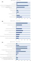
Data collection and analysis: Two reviewers and a statistician reviewed study design, statistical methods, and reporting methods using 29 criteria. Each article was awarded an overall quality score, predefined as the percentage of the 29 criteria scored at low risk of bias. 95% prediction intervals were calculated for studies that used recommended first trimester dating to confirm true GA.
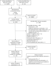
Main results: The search yielded 4209 results. Ninety-seven full-text articles were included in the analysis. The mean quality score was 32% (range 7%-97%). Only 10 articles scored low risk in 18 or more criteria. Their formulas estimated GA using one or more ultrasound-measured biometry parameters and SFH measurements. Twenty-three articles used recommended first trimester dating. A single-parameter formula using transcerebellar diameter (TCD) gave the lowest 95% prediction interval.
Conclusions: There is considerable methodological heterogeneity in studies developing equations for estimating GA. Formulas using ultrasound-based measurements more accurately estimated GA after 20 weeks than formulas using SFH measurement. While the clinical priority remains promotion of early engagement with antenatal care, we suggest unified standards for GA and growth assessment.
Tweetable abstract: Many vulnerable women seek antenatal care late in pregnancy. How should gestational age be determined? We examine all available studies estimating GA >20 weeks. Ultrasound is much better than fundal height, and using cerebellar measurement appears to be the most accurate.
Keywords: biometry; due date; gestational age; growth; ltrasound dating; post-term; pregnancy; pregnancy dating; preterm; screening.
Conflict of interest statement
Completed disclosure of interest forms are available to view online as supporting information.
Figures
Comment in
-
The value of fetal size measurement in the estimation of gestational age in low and middle income countries.BJOG. 2022 Aug;129(9):1459. doi: 10.1111/1471-0528.17127. Epub 2022 Apr 3. BJOG. 2022. PMID: 35195328 No abstract available.
References
-
- Campbell S, Warsof SL, Little D, Cooper DJ. Routine ultrasound screening for the prediction of gestational age. Obstet Gynecol. 1985;65(5):613–20. - PubMed
-
- Mukri F, Bourne T, Bottomley C, Schoeb C, Kirk E, Papageorghiou AT. Evidence of early first‐trimester growth restriction in pregnancies that subsequently end in miscarriage. BJOG. 2008;115(10):1273–8. - PubMed
-
- Butt K, Lim KI. Guideline no. 388‐determination of gestational age by ultrasound. J Obstet Gynaecol Can. 2019;41(10):1497–507. - PubMed
-
- Salomon LJ, Alfirevic Z, Bilardo CM, Chalouhi GE, Ghi T, Kagan KO, et al. ISUOG practice guidelines: performance of first‐trimester fetal ultrasound scan. Ultrasound Obstet Gynecol. 2013;41(1):102–13. - PubMed
Publication types
MeSH terms
Grants and funding
LinkOut - more resources
Full Text Sources
Miscellaneous


