. 2022 Mar 14;8(3):1239-1246.
doi: 10.1021/acsbiomaterials.1c01163. Epub 2022 Feb 14.

Impact of Membrane Voltage on Formation and Stability of Human Renal Proximal Tubules in Vitro

Affiliations

Impact of Membrane Voltage on Formation and Stability of Human Renal Proximal Tubules in Vitro

Miryam Adelfio et al. ACS Biomater Sci Eng. .

Abstract

More than 15% of adults in the United States suffer from some form of chronic kidney disease (CKD). Current strategies for CKD consist of dialysis or kidney transplant, which, however, can take several years. In this light, tissue engineering and regenerative medicine approaches are the key to improving people's living conditions by advancing previous tissue engineering approaches and seeking new targets as intervention methods for kidney repair or replacement. The membrane voltage (Vm) dynamics of a cell have been associated with cell migration, cell cycle progression, differentiation, and pattern formation. Furthermore, bioelectrical stimuli have been used as a means in the treatment of diseases and wound healing. Here, we investigated the role of Vm as a novel target to guide and manipulate in vitro renal tissue models. Human-immortalized renal proximal tubule epithelial cells (RPTECs-TERT1) were cultured on Matrigel to support the formation of 3D proximal tubular-like structures with the incorporation of a voltage-sensitive dye indicator─bis-(1,3-dibutylbarbituric acid)timethine oxonol (DiBAC). The results demonstrated a correlation between the depolarization and the reorganization of human renal proximal tubule cells, indicating Vm as a candidate variable to control these events. Accordingly, Vm was pharmacologically manipulated using glibenclamide and pinacidil, KATP channel modulators, and proximal tubule formation and tubule stability over 21 days were assessed. Chronic manipulation of KATP channels induced changes in the tubular network topology without affecting lumen formation. Thus, a relationship was found between the preluminal tubulogenesis phase and KATP channels. This relationship may provide future options as a control point during kidney tissue development, treatment, and regeneration goals.

Keywords: kidney; membrane potential; proximal; tubulogenesis; voltage.


Conflict of interest statement

Competing interests

The authors declare no competing financial interest.

Figures

Figure 1.
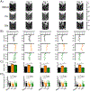
Figure 1.. Vm trajectory during cell reorganization and KATP electrophysiological properties in RPTECs.
A) Representative heatmap images of normalized DiBAC signal. Scale bar: 100 μm. B) Percentage of MFI intensity trajectory normalized on day 1 (top) and ΔV voltage trajectory compared to day 1 (bottom). C) Voltage protocol (top, blue; holding potential (hp)=−60 mV, from −120 mV to 40 mV, 400 ms duration) and representative current traces (bottom) in DMSO (green) and glibenclamide (red) conditions; the glibenclamide-sensitive current trace is shown in black. D) The amount of IKATP current at the holding potential before and after application of glibenclamide in the bath (grey circles); mean IKATP currents at the holding potential in DMSO and glibenclamide (black squares). On the right, the mean ERev identified as the voltage at which the current flow was zero (indicated with an arrow in panel E). E) Mean glibenclamide-sensitive current density-voltage plot. *p=0.005. Paired T-Students test. Data shown as Mean ± S.E.M.
Figure 2.
Figure 2.. The Vm trajectory was not affected by chronic manipulation of the KATP channels.
A) Representative heatmap images of normalized DiBAC signal for the different conditions and days, as indicated. Scale bar: 100 μm. B) Single MFI values (circles) and mean MFI intensities (bar graphs) for DMSO, pinacidil and glibenclamide (black, orange, and green, respectively). Single logarithmic ratios (pinacidil/DMSO and glibenclamide/DMSO; diamonds) and mean logarithmic ratios (box plots) for the two treatments. Number of experiments per day: day 1 DMSO (8), pinacidil (5) and glibenclamide (7); day 3 DMSO (6), pinacidil (3) and glibenclamide (3); day 7 DMSO (8), pinacidil (4) and glibenclamide (4); day 14 DMSO (8), pinacidil (4) and glibenclamide (5); day 21 DMSO (7), pinacidil (4) and glibenclamide (4).
Figure 3.
Figure 3.. The KATP channels shape density and length of tubular structures.
A) Representative tubular formation for the different conditions and days, as indicated. B) Number of intersections/mm (x-axis) as a function of the circumference values (y-axis) of the circles used for the Sholl analysis. C) Mean intersections/mm and D) tubule lengths for the different conditions and days, as indicated. *p<0.05, One-Way ANOVA, Fisher post-hoc test. Number of experiment 3 and number of tubules analyzed 66/3 images.

Similar articles

Cited by

References

    1. Levin M, Pezzulo G, Finkelstein JM. Endogenous Bioelectric Signaling Networks: Exploiting Voltage Gradients for Control of Growth and Form. Annu Rev Biomed Eng. 2017;19:353–87. - PMC - PubMed
    1. Levin M Bioelectric signaling: Reprogrammable circuits underlying embryogenesis, regeneration, and cancer. Cell. 2021. - PubMed
    1. Hille B Ion channels of excitable membranes. 3rd ed. Sunderland, Mass.: Sinauer; 2001. xviii, 814
    1. Abdul Kadir L, Stacey M, Barrett-Jolley R. Emerging Roles of the Membrane Potential: Action Beyond the Action Potential. Front Physiol. 2018;9:1661. - PMC - PubMed
    1. A Meta-Analysis of Bioelectric Data in Cancer, Embryogenesis, and Regeneration.0(0):null. - PMC - PubMed

Publication types

Substances

LinkOut - more resources