

Hotspot analysis by confocal microscopy can help to differentiate challenging melanocytic skin lesions
- PMID: 35157706
- PMCID: PMC8843198
- DOI: 10.1371/journal.pone.0263819
Hotspot analysis by confocal microscopy can help to differentiate challenging melanocytic skin lesions
Abstract
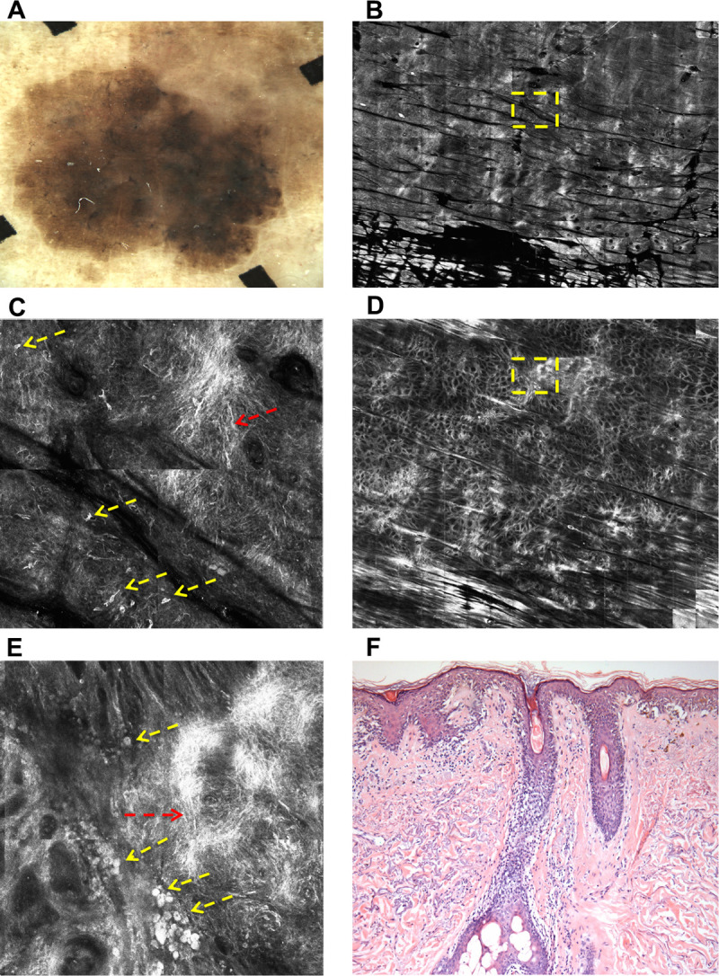
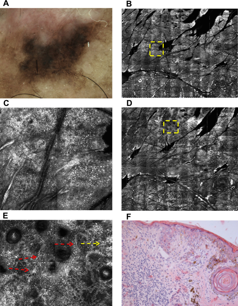
Some melanocytic lesions do not present enough clinical and dermoscopic features to allow ruling out a possible melanoma diagnosis. These "doubtful melanocytic lesions" pose a very common and challenging scenario in clinical practice and were selected at this study for reflectance confocal microscopy evaluation and subsequent surgical excision for histopathological diagnosis. The study included 110 lesions and three confocal features were statistically able to distinguish benign melanocytic lesions from melanomas: "peripheral hotspot at dermo-epidermal junction", "nucleated roundish cells at the dermo-epidermal junction" and "sheet of cells". The finding of a peripheral hotspot (atypical cells in 1mm2) at the DEJ is highlighted because has not been previously reported in the literature as a confocal feature related to melanomas.
Conflict of interest statement
The authors have declared that no competing interests exist.
Figures
Similar articles
-
Role of In Vivo Reflectance Confocal Microscopy in the Analysis of Melanocytic Lesions.Acta Dermatovenerol Croat. 2018 Apr;26(1):64-67. Acta Dermatovenerol Croat. 2018. PMID: 29782304 Review.
-
Reflectance confocal microscopy analysis of equivocal melanocytic lesions with severe regression.Skin Res Technol. 2018 Feb;24(1):9-15. doi: 10.1111/srt.12382. Epub 2017 May 21. Skin Res Technol. 2018. PMID: 28543606
-
Pigmented skin lesions displaying regression features: Dermoscopy and reflectance confocal microscopy criteria for diagnosis.Exp Dermatol. 2019 Feb;28(2):129-135. doi: 10.1111/exd.13853. Epub 2019 Jan 14. Exp Dermatol. 2019. PMID: 30506970
-
Dendritic cells in reflectance confocal microscopy are a clue for early melanoma diagnosis in extrafacial flat pigmented melanocytic lesions.Exp Dermatol. 2022 Jul;31(7):1048-1055. doi: 10.1111/exd.14553. Epub 2022 Mar 4. Exp Dermatol. 2022. PMID: 35220636 Free PMC article.
-
Discriminating Nevi from Melanomas: Clues and Pitfalls.Dermatol Clin. 2016 Oct;34(4):395-409. doi: 10.1016/j.det.2016.05.003. Dermatol Clin. 2016. PMID: 27692446 Free PMC article. Review.
References
MeSH terms
LinkOut - more resources
Full Text Sources
Medical
