Review
. 2022 Jul;77(1):206-218.
doi: 10.1016/j.jhep.2022.01.025. Epub 2022 Feb 11.

Hepatic lymphatic vascular system in health and disease

Affiliations
Review

Hepatic lymphatic vascular system in health and disease

Jain Jeong et al. J Hepatol. 2022 Jul.

Abstract

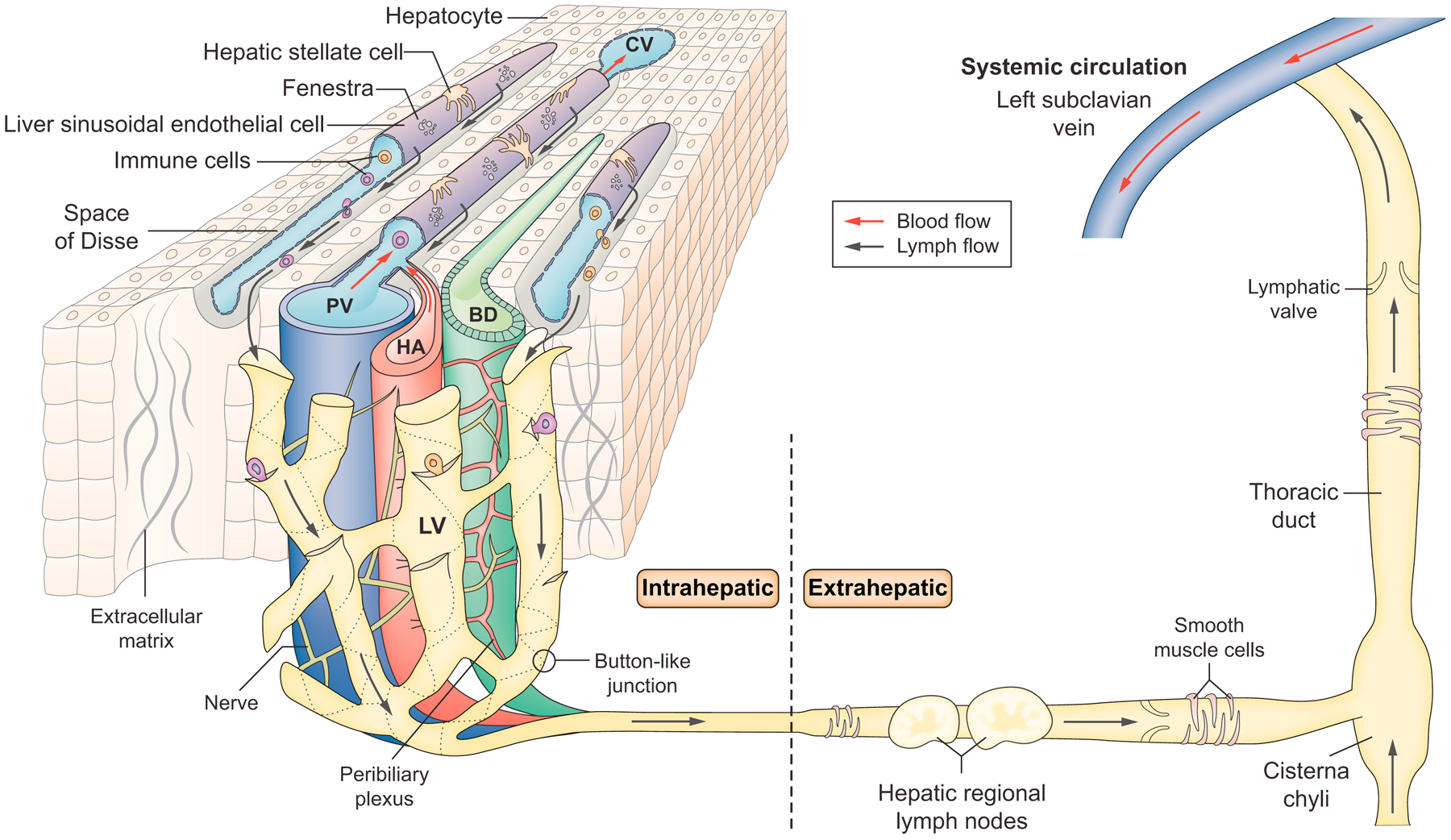
In recent years, significant advances have been made in the study of lymphatic vessels with the identification of their specific markers and the development of research tools that have accelerated our understanding of their role in tissue homeostasis and disease pathogenesis in many organs. Compared to other organs, the lymphatic system in the liver is understudied despite its obvious importance for hepatic physiology and pathophysiology. In this review, we describe fundamental aspects of the hepatic lymphatic system and its role in a range of liver-related pathological conditions such as portal hypertension, ascites formation, malignant tumours, liver transplantation, congenital liver diseases, non-alcoholic fatty liver disease, and hepatic encephalopathy. The article concludes with a discussion regarding the modulation of lymphangiogenesis as a potential therapeutic strategy for liver diseases.

Keywords: Lymphangiogenesis; VEGFs; liver fibrosis; liver transplantation; portal hypertension.


Conflict of interest statement

Conflict of interest The authors declare no conflicts of interest that pertain to this work. Please refer to the accompanying ICMJE disclosure forms for further details.

Figures

Figure 1
Figure 1. Hepatic lymphatic system
Lymph is thought to flow into lymphatic vessels located in three regions: portal, hepatic venous and sub-capsular areas. The illustration shows lymphatic vessels in the portal tract, the primary site of hepatic lymph drainage covering around 80% of the lymph produced by the liver. Because the hepatic sinusoids are highly permeable due to fenestrae, fluid in the hepatic sinusoids can flow through the channels traversing the limiting plate to the interstitial space of the portal tract. Endothelial cells of lymphatic capillaries have discontinuous, “button-like” junctions, which allow efficient entry of fluid, antigens and immune cells into lymphatic capillaries. Lymphatic capillaries in the portal tract coalesce into collecting lymphatic vessels surrounded by lymphatic muscle cells outside the liver. Lymphatic muscle cells covering collecting lymphatic vessels help to pump lymph into regional lymph nodes located in the hepatic hilum and then to the cisterna chyli located at the lower end of the thoracic duct. Lymph finally drains into the left subclavian vein via the thoracic duct and returns to the systemic blood circulation. LV: Lymphatic vessel, PV: Portal vein, HA: Hepatic artery, BD: Bile duct, CV: Central vein, ECM: Extracellular matrix protein, HSC: Hepatic stellate cell
Figure 2
Figure 2. Button vs. Zipper-like structures
The lymphatic vascular system includes lymphatic capillaries (also known as initial lymphatics) and collecting lymphatic vessels. Lymphatic capillaries consist of a single layer of lymphatic endothelial cells. Endothelial cells of lymphatic capillaries are attached by anchoring filaments to surrounding extracellular matrix proteins, which support their vessel structure. Endothelial cells of lymphatic capillaries have discontinuous, “button-like” junctions, which allow efficient entry of fluid, antigens and immune cells into lymphatic capillaries. In contrast, collecting lymphatic vessels have “zipper-like” junctions, similar to blood vessels. In pathological conditions, lymphatic capillaries lose this “button-like” structure with a change to the less permeable “zipper-like” structure, which results in impaired transport of fluid and substances to lymphatic capillaries, thereby decreasing their clearance from tissues. Created with BioRender.com.
Figure 3
Figure 3. VEGFs and VEGFRs in angiogenesis and lymphangiogenesis
VEGF (Vascular endothelial growth factor) is a potent mediator of both angiogenesis and lymphangiogenesis. All members of the VEGF family induce cellular responses by binding to specific VEGF receptors with tyrosine kinases, leading them to dimerize and activate through phosphorylation. VEGF-C and D bind strongly to VEGFR3/Flt4 and induce lymphangiogenesis, while they also bind very weakly to VEGFR2/KDR. VEGF-A binds to VEGFR1/Flt1 and VEGFR2/KDR and mediates angiogenesis. VEGF-B and placental growth factor (PIGF) bind only to VEGFR1/Flt1. Thick and thin arrows indicate strong and weak binding, respectively. Created with BioRender.com.
Figure 4
Figure 4. Mechanisms of ascites formation in liver cirrhosis with portal hypertension
Elevated hydrostatic pressure in the sinusoids due to liver cirrhosis causes an increased production of lymph. It is thought that ascitic fluid starts to accumulate when capsular or superficial lymphatics of the liver rupture and hepatic lymph with a high protein concentration leaks into the peritoneal cavity. This lymph leakage from the surface of the liver is known as the so-called ‘weeping liver’. When the total fluid flux into the peritoneal cavity exceeds the lymph draining capacity of the peritoneum, ascites forms. According to the “forward theory”, portal hypertension causes excessive arterial vasodilation in the splanchnic arterial circulation, leading to the underfilling of the arterial circulation or hypovolemia. Splanchnic arterial vasodilation makes sodium and water retention persistent and leads to leaking fluid into the peritoneal cavity and accumulation of ascites. Created with BioRender.com.

References

    1. Morris B The hepatic and intestinal contributions to the thoracic duct lymph. Q J Exp Physiol Cogn Med Sci 1956;41:318–325. - PubMed
    1. Cain JC, Grindlay JH, et al. Lymph from liver and thoracic duct; an experimental study. Surg Gynecol Obstet 1947;85:558–562. - PubMed
    1. Mobley WP, Kintner K, Witte CL, Witte MH. Contribution of the liver to thoracic duct lymph flow in a motionless subject. Lymphology 1989;22:81–84. - PubMed
    1. Tanaka M, Iwakiri Y. The Hepatic Lymphatic Vascular System: Structure, Function, Markers, and Lymphangiogenesis. Cell Mol Gastroenterol Hepatol 2016;2:733–749. - PMC - PubMed
    1. Ohtani O, Ohtani Y. Lymph circulation in the liver. Anat Rec (Hoboken) 2008;291:643–652. - PubMed

Publication types