. 2022 Jan 28;14(3):673.
doi: 10.3390/cancers14030673.

Dual Inhibition of Myc Transcription and PI3K Activity Effectively Targets Colorectal Cancer Stem Cells

Affiliations

Dual Inhibition of Myc Transcription and PI3K Activity Effectively Targets Colorectal Cancer Stem Cells

Miriam Gaggianesi et al. Cancers (Basel). .

Abstract

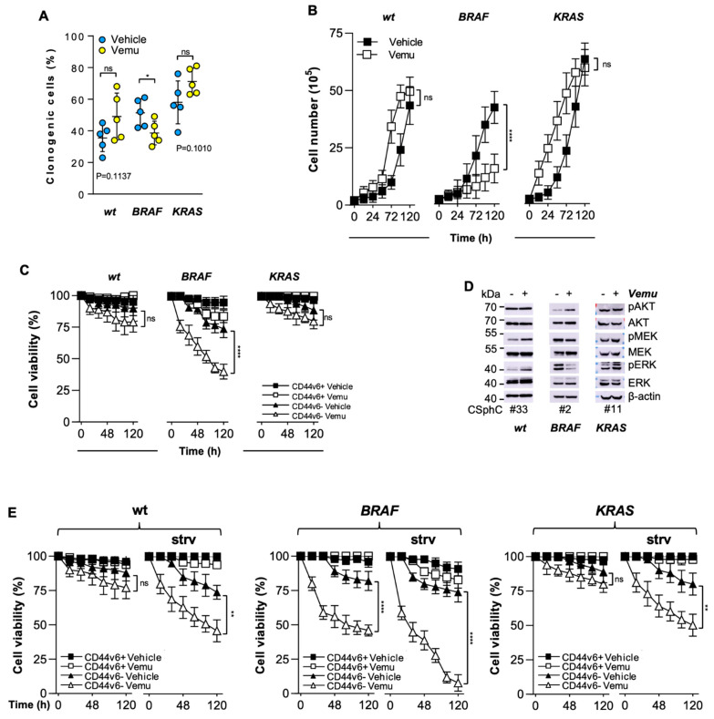
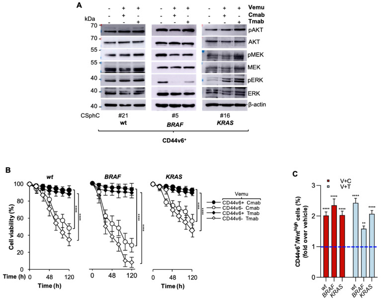
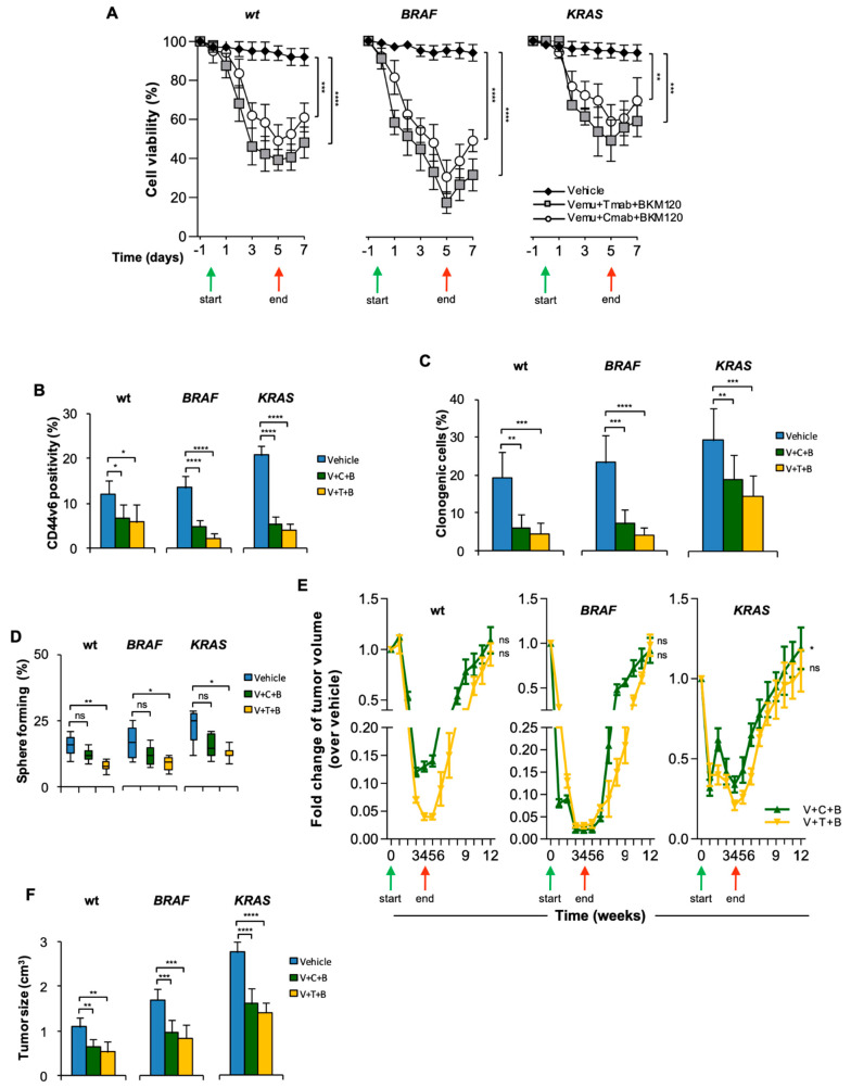
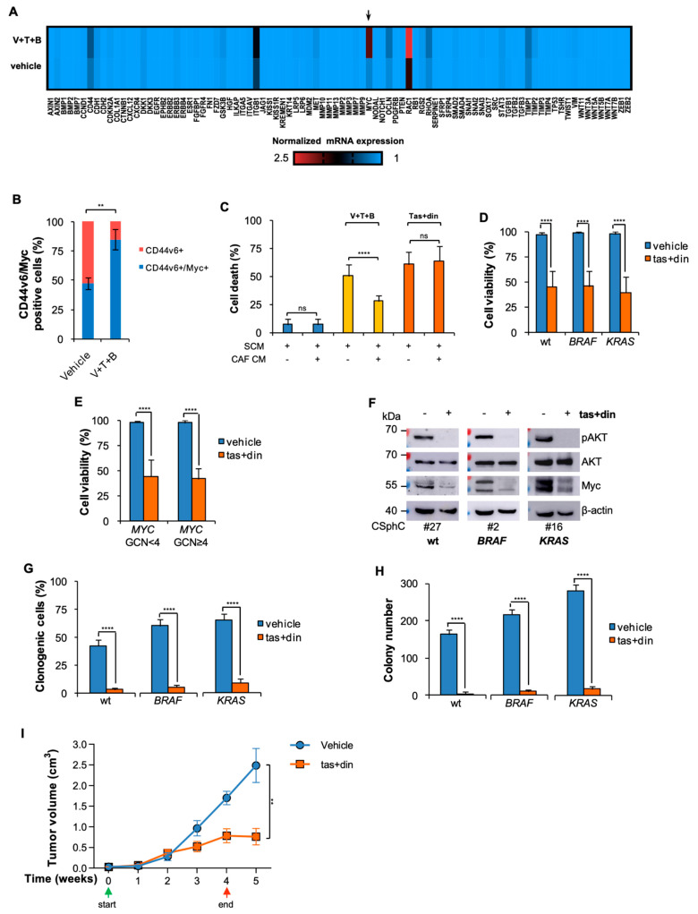
Despite advances in the curative approach, the survival rate of advanced colorectal cancer (CRC) patients is still poor, which is likely due to the emergence of cancer cell clones resistant to the available therapeutic options. We have already shown that CD44v6-positive CRC stem cells (CR-CSCs) are refractory toward standard anti-tumor therapeutic agents due to the activation of the PI3K pathway together with high HER2 expression levels. Tumor microenvironmental cytokines confer resistance to CR-CSCs against HER2/PI3K targeting by enhancing activation of the MAPK pathway. Here, we show that the CSC compartment, spared by BRAF inhibitor-based targeted therapy, is associated with increased expression levels of CD44v6 and Myc and retains boosted clonogenic activity along with residual tumorigenic potential. Inhibition of Myc transcription, downstream of the MAPK cascade components, and PI3K pathway activity was able to overcome the protective effects of microenvironmental cytokines, affecting the survival and the clonogenic activity of CR-CSCs, regardless of their mutational background. Likewise, the double targeting induced stabilization of mouse tumor avatars. Altogether, these data outline the rationale for dual kinase targeting of CR-CSCs to prevent their adaptive response, which would lead to disease progression.

Keywords: anti-tumor drug resistance; cancer stem cells; colorectal cancer; combination therapies.


Conflict of interest statement

The authors declare no conflict of interest.

Figures

Figure 1
Figure 1
The resistance of CD44v6-positive CR-CSCs to BRAF inhibition is mediated by EGFR-driven MAPK pathway activation. (A) Clonogenic assays for wt (#6, #14, #21, #27, #33), BRAF- (#1, #2, #3, #4, #5) and KRAS-mutant (#9, #10, #11, #13, #16) CSphCs pretreated with a vehicle (Vehicle) or vemurafenib (Vemu, 1 µM) for 120 h. The statistical significance between two groups was determined with a paired two-tailed Student’s t-test. (B) Kinetics of wt (#14, 21), BRAF- (#2, 5) and KRAS-mutant (#11, 16) sphere cell growth following treatment with vemurafenib (Vemu, 1 µM) for up to 120 h. (C) Cell viability percentage of CD44v6+ and CD44v6 wt (#21, #27, #33), BRAF- (#1, #3, #5) and KRAS-mutant (#9, #13, #16) sphere cells treated as in (B). (D) Immunoblot analysis of pAKT, AKT, pMEK, MEK, pERK and ERK on the indicated CR-CSphCs treated with a vehicle (−) or vemurafenib (Vemu, +, 1 µM) for 48 h. (E) Cell viability percentage of CD44v6+ and CD44v6 wt (#21, #27, #33), BRAF- (#1, #3, #5) and KRAS-mutant (#9, #13, #16) sphere cells treated as indicated in the presence or absence (strv) of EGF for up to 120 h. For (BC,E), data are means ± SD from three independent experiments. ns, not significant; * p ≤ 0.05; ** p ≤ 0.01, **** p ≤ 0.0001. The uncropped blots are shown in supplementary materials.
Figure 2
Figure 2
PI3K activation protects CD44v6-positive cells from EGFR/HER2 blockade in combination with BRAF inhibition. (A) Immunoblot of pAKT, AKT, pMEK, MEK, pERK and ERK on the indicated CD44v6+ cells exposed to a vehicle (−) or to vemurafenib (Vemu, +, 1 µM) in combination with cetuximab (Cmab, +, 20 µg/mL) or trastuzumab (Tmab, +, 10 µg/mL) for 2 h. (B) Cell viability percentage in CD44v6+ and CD44v6 wt (#21, #27), BRAF- (#1, #5) and KRAS-mutant (#11, #16) CR-CSphCs treated with vemurafenib (Vemu, 1 µM) in combination with cetuximab (Cmab, 20 µg/mL) or trastuzumab (Tmab, 10 µg/mL). Data are shown as means ± SD of three independent experiments for each CR-CSphC line. (C) Fold increase over vehicle (blue dotted line) of CD44v6+/Wnthigh subpopulation in wt (#27), BRAF- (#1) and KRAS-mutant (#9) TOP-GFP transduced sphere cells, treated as indicated for 120 h. Data represent the means ± SD of three independent experiments. ** p ≤ 0.01, **** p ≤ 0.0001.The uncropped blots are shown in supplementary materials.
Figure 3
Figure 3
HER2/BRAF/PI3K combinatorial targeting leads to transient therapeutic response. (A) Cell viability in wt (#21, #27, #33), BRAF- (#1, #3, #5) and KRAS-mutant (# 9, #11, #16) CR-CSphCs treated with Vemu (1 µM) + Tmab (10 µg/mL) + BKM120 (1 µM), Vemu + Cmab (20 µg/mL) + BKM120 or with a vehicle as control. (B) Percentage of CD44v6 positivity evaluated by flow cytometry for wt (#21, #27, #33), BRAF- (#1, #3, #5) and KRAS-mutant (#9, #11, #16) CR-CSphCs treated as indicated for 72 h. (C,D) Clonogenic activity (C) and percentage of sphere-forming cells for wt (#21, #27, #33), BRAF- (#1, #3, #5) and KRAS-mutant (#9, #11, #16) sphere cells pretreated as in (B). For (AD), data show the means ± SD of three different experiments. (E) Fold change over vehicle of volume of subcutaneous tumor xenograft generated by the injection of wt (#14, 21), BRAF- (#1, 2) or KRAS-mutant (#11, 16) CR-CSphCs and treated with a vehicle (Vehicle), vemurafenib (V, 20 mg/Kg), cetuximab (C, 40 mg/Kg) or trastuzumab (T, 5 mg/Kg) in combination with BKM120 (B, 20 mg/Kg) for 4 weeks. For (A,E), green and red arrows indicate the start and the end of the treatment, respectively. (F) Size of secondary tumors (8 weeks) generated by subcutaneous injection of 1 × 10⁴ freshly purified CR-CSphCs derived from wt (#21), BRAF- (#2) or KRAS-mutant (#11) xenograft tumors. For (D,E), data are expressed as means ± SD (n = 6). ns, not significant; * p ≤ 0.05, ** p ≤ 0.01, *** p ≤ 0.001, **** p ≤ 0.0001.
Figure 4
Figure 4
Myc and PI3K combinatorial targeting reduces CR-CSphC viability, downregulating Myc and CD44v6 expression levels. (A) Heatmap of cancer stem cell- and metastasis-related genes (2−ΔCt) in freshly purified cells (#21, #2, #10) from xenografts treated with a vehicle or V (20 mg/Kg) + T (5 mg/Kg) + B (20 mg/Kg). (B) Percentage of CD44v6 and Myc expression in xenograft tumors treated for 4 weeks with a vehicle (Vehicle) or V (20 mg/Kg) + T (5 mg/Kg) + B (20 mg/Kg). Data are shown as means ± SD of three different xenografts (#21, #2, #11). (C) Death percentage of CR-CSphCs treated as indicated in the presence of SCM or CAF-conditioned medium (CAF CM) for 72 h. Data represent means ± SD of three independent experiments performed with wt (#21, #27, #33), BRAF- (#1, #3, #5) and KRAS-mutant (#8, #9, #16) CR-CSphCs. (D) Cell viability in wt (#6, #14, #21, #27, #33, #49), BRAF- (#1–#5) and KRAS-mutant (#8, #9, #11, #13, #16, #59) CR-CSphCs treated with vehicle or taselisib (1 µM) and dinaciclib (10 nM) (tas + din) for 72 h. (E) Cell viability in MYC wt (MYC GCN < 4, CSphC#2, #3, #6, #9, #14, #16, #21, #27, #33) and MYC-amplified (MYC GCN ≥ 4, #1, #4, #5, #8, #11, #13, #49, #59) CR-CSphCs treated as in (D). For (C,D), data represent means ± SD of three independent experiments. (F) Analysis of pAKT, AKT and Myc on the indicated CR-CSphCs and treated with a vehicle or tas (1 µM) + din (10 nM) at 48 h. (G,H) Clonogenic activity (G) and colony number (H) of wt (#21, #33), BRAF- (#1, #3) and KRAS-mutant (#9, #16) sphere cells previously treated with a vehicle or tas (1 µM) + din (10 nM) for 72 h, in the presence of CAF CM. For (G,H), data are means ± SD of three independent experiments. For (DH), CR-CSphCs were exposed to CAF CM. (I) Tumor volume of subcutaneous xenograft generated by the injection of KRAS-mutant (#8) CR-CSphCs and treated with a vehicle (Vehicle) or taselisib (tas, 5 mg/Kg) and dinaciclib (din, 25 mg/Kg) for 4 weeks. Green and red arrows indicate the start and the end of the treatment, respectively. Data are expressed as means ± SD (n = 6). ns, not significant; ** p ≤ 0.01, **** p ≤ 0.0001. The uncropped blots are shown in supplementary materials.

References

    1. Siegel R.L., Miller K.D., Fuchs H.E., Jemal A. Cancer Statistics, 2021. CA Cancer J. Clin. 2021;71:7–33. doi: 10.3322/caac.21654. - DOI - PubMed
    1. Xie Y.-H., Chen Y.-X., Fang J.-Y. Comprehensive review of targeted therapy for colorectal cancer. Signal Transduct. Target. Ther. 2020;5:1–30. doi: 10.1038/s41392-020-0116-z. - DOI - PMC - PubMed
    1. Raskov H., Søby J.H., Troelsen J.T., Bojesen R.D., Gögenur I. Driver Gene Mutations and Epigenetics in Colorectal Cancer. Ann. Surg. 2020;271:75–85. doi: 10.1097/SLA.0000000000003393. - DOI - PubMed
    1. Matano M., Date S., Shimokawa M., Takano A., Fujii M., Ohta Y., Watanabe T., Kanai T., Sato T. Modeling colorectal cancer using CRISPR-Cas9–mediated engineering of human intestinal organoids. Nat. Med. 2015;21:256–262. doi: 10.1038/nm.3802. - DOI - PubMed
    1. Gaggianesi M., Di Franco S., Pantina V.D., Porcelli G., D’Accardo C., Verona F., Veschi V., Colarossi L., Faldetta N., Pistone G., et al. Messing Up the Cancer Stem Cell Chemoresistance Mechanisms Supported by Tumor Microenvironment. Front. Oncol. 2021;11:2847. doi: 10.3389/fonc.2021.702642. - DOI - PMC - PubMed