Spatial Immunology in Liver Metastases from Colorectal Carcinoma according to the Histologic Growth Pattern
- PMID: 35158957
- PMCID: PMC8833601
- DOI: 10.3390/cancers14030689
Spatial Immunology in Liver Metastases from Colorectal Carcinoma according to the Histologic Growth Pattern
Abstract
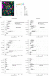
Colorectal cancer liver metastases (CRC-LM) present differential histologic growth patterns (HGP) that determine the interaction between immune and tumor cells. We explored the spatial distribution of lymphocytic infiltrates in CRC-LM in the context of the HGP using multispectral digital pathology. We did not find statistically significant differences of immune cell densities in the central regions of desmoplastic (dHGP) and non-desmoplastic (ndHGP) metastases. The spatial evaluation reported that dHGP-metastases displayed higher infiltration by CD8+ and CD20+ cells in peripheral regions as well as CD4+ and CD45RO+ cells in ndHGP-metastases. However, the reactive stroma regions at the invasive margin (IM) of ndHGP-metastases displayed higher density of CD4+, CD20+, and CD45RO+ cells. The antitumor status of the TIL infiltrates measured as CD8/CD4 reported higher values in the IM of encapsulated metastases up to 400 μm towards the tumor center (p < 0.05). Remarkably, the IM of dHGP-metastases was characterized by higher infiltration of CD8+ cells in the epithelial compartment parameter assessed with the ratio CD8epithelial/CD8stromal, suggesting anti-tumoral activity in the encapsulating lesions. Taking together, the amount of CD8+ cells is comparable in the IM of both HGP metastases types. However, in dHGP-metastases some cytotoxic cells reach the tumor nests while remaining retained in the stromal areas in ndHGP-metastases.
Keywords: desmoplasia; growth pattern; immunology; liver metastases; lymphocyte; multiplex.
Conflict of interest statement
None of the authors have any conflict of interest to declare.
Figures


References
-
- Adam R., De Gramont A., Figueras J., Guthrie A., Kokudo N., Kunstlinger F., Loyer E., Poston G., Rougier P., Rubbia-Brandt L., et al. The oncosurgery approach to managing liver metastases from colorectal cancer: A multidisciplinary international consensus. Oncologist. 2012;17:1225–1239. doi: 10.1634/theoncologist.2012-0121. - DOI - PMC - PubMed
-
- Van Cutsem E., Tabernero J., Lakomy R., Prenen H., Prausová J., Macarulla T., Ruff P., Van Hazel G.A., Moiseyenko V., Ferry D., et al. Addition of aflibercept to fluorouracil, leucovorin, and irinotecan improves survival in a phase III randomized trial in patients with metastatic colorectal cancer previously treated with an oxaliplatin-based regimen. J. Clin. Oncol. 2012;30:3499–3506. doi: 10.1200/JCO.2012.42.8201. - DOI - PubMed
-
- Johnsson A., Hagman H., Frodin J.E., Berglund Å., Keldsen N., Fernebro E., Sundberg J., Christensen R.D.P., Spindler K.L.G., Bergström D., et al. A randomized phase III trial on maintenance treatment with bevacizumab alone or in combination with erlotinib after chemotherapy and bevacizumab in metastatic colorectal cancer: The Nordic ACT Trial. Ann. Oncol. 2013;24:2335–2341. doi: 10.1093/annonc/mdt236. - DOI - PubMed
Grants and funding
LinkOut - more resources
Full Text Sources
Research Materials

