. 2022 Jan 30;14(3):716.
doi: 10.3390/cancers14030716.

MRI-Guided Online Adaptive Stereotactic Body Radiation Therapy of Liver and Pancreas Tumors on an MR-Linac System

Affiliations

MRI-Guided Online Adaptive Stereotactic Body Radiation Therapy of Liver and Pancreas Tumors on an MR-Linac System

Teo Stanescu et al. Cancers (Basel). .

Abstract

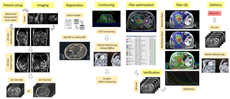
Purpose: To describe a comprehensive workflow for MRI-guided online adaptive stereotactic body radiation therapy (SBRT) specific to upper gastrointestinal cancer patients with abdominal compression on a 1.5T MR-Linac system. Additionally, we discuss the workflow's clinical feasibility and early experience in the case of 16 liver and pancreas patients.

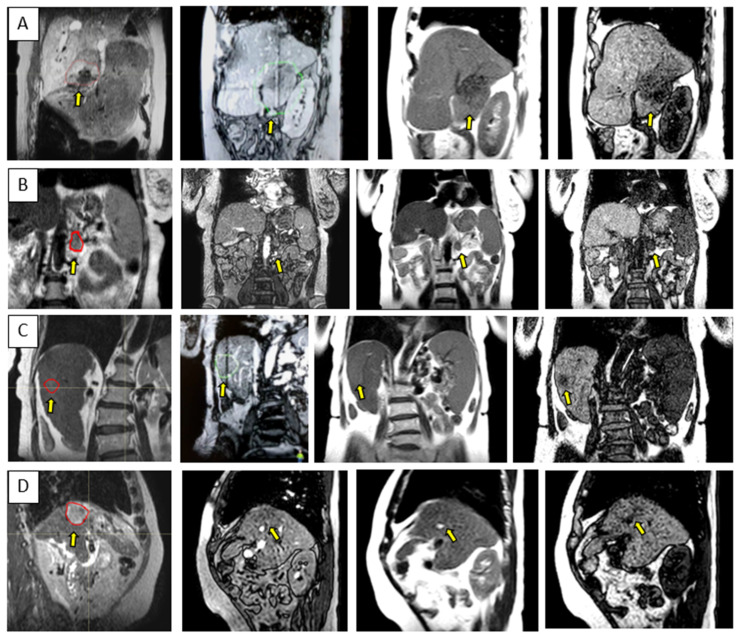
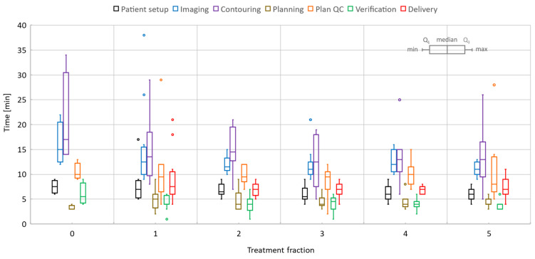
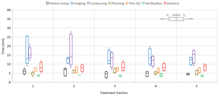
Methods: Eleven patients with liver cancer and five patients with pancreas cancer were treated with online adaptive MRI-guidance under abdominal compression. Two liver patients received single-fraction treatments; the remainder plus all pancreas cancer patients received five fractions. A total of 65 treatment sessions were investigated to provide analytics relevant to the online adaptive processes. The quantification of target and organ motion as well as definition and validation of internal target volume (ITV) margins were performed via multi-contrast imaging provided by three different 2D cine sequences. The plan generation was driven by full re-optimization strategies and using T2-weighted 3D image series acquired by means of a respiratory-triggered exhale phase or a time-averaged imaging protocol. As a pre-requisite for the clinical development of the procedure, the image quality was thoroughly investigated via phantom measurements and numerical simulations specific to upper abdominal sites. The delivery of the online adaptive treatments was facilitated by real-time monitoring with 2D cine imaging.

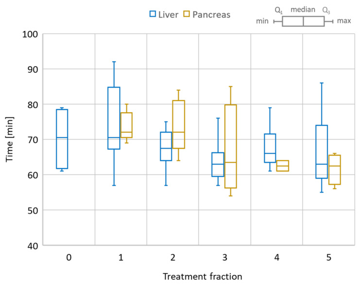
Results: Liver 1-fraction and 5-fraction online adaptive session time were on average 80 and 67.5 min, respectively. The total session length varied between 70-90 min for a single fraction and 55-90 min for five fractions. The pancreas sessions were 54-85 min long with an average session time of 68.2 min. Target visualization on the 2D cine image data varied per patient, with at least one of the 2D cine sequences providing sufficient contrast to confidently identify its location and confirm reproducibility of ITV margins. The mean/range of absolute and relative dose values for all treatment sessions evaluated with ArcCheck were 90.6/80.9-96.1% and 99/95.4-100%, respectively.

Conclusion: MR-guidance is feasible for liver and pancreas tumors when abdominal compression is used to reduce organ motion, improve imaging quality, and achieve a robust intra- and inter-fraction patient setup. However, the treatment length is significantly longer than for the conventional linac, and patient compliance is paramount for the successful completion of the treatment. Opportunities for reducing the online adaptive session time should be explored. As the next steps, dose-of-the-day and dose accumulation analysis and tools are needed to enhance the workflow and to help further refine the online re-planning processes.

Keywords: HCC; MR image distortions; MR-Linac; MR-guided radiation therapy; MRI; MRIgRT; MRgRT; SBRT; liver metastases; numerical simulations; pancreas; susceptibility.


Conflict of interest statement

A.H.—Non-financial leadership of the liver tumor site group at Elekta MRL Consortium. The rest of the authors declare no conflict of interest.

Figures

Figure 1
Figure 1
Flow chart highlighting the key processes in the MRI-guided online adaptive workflow based on the adapt-to-shape (ATS) procedure for UGI sites on the Unity MR-Linac system.
Figure 2
Figure 2
Representation of the MR image distortion fields relevant to liver RT and imaging performed on the Unity-MR system: (A) 3D mesh of the liver volume used to plot the distortion field distribution; (B) total distortion values; (C) system-related spatial distortions; (D) susceptibility-induced geometric distortions.
Figure 3
Figure 3
Liver examples depicting the comparison between the T2w-Nav (first column) and T2w-Ave (second column) image sequences. The target(s) location is identified by the yellow arrow. The samples correspond to Patients 7, 8, and 9 as per Table 1. These cases were planned with the T2w-Nav image data.
Figure 4
Figure 4
Pancreas examples highlighting the comparison between the T2w-Nav (first column) and T2w-Ave (second column) image sequences. The target location is identified by the yellow dashed circle. The planning data set was the T2w-Nav for the first case and T2w-Ave for the second and third case. The samples correspond to Patients 11, 14, and 16 as listed in Table 1.
Figure 5
Figure 5
Sample data for four UGI patients highlighting the range of contrast available with the 3D cine sequences used for the quantification and monitoring of patient setup and target motion. Each row is for a different patient and shows either a coronal or sagittal view: (AD) corresponds to Patients 10, 15, 6, and 12, as per Table 1. The columns correspond from left to right to the MR planning data set identifying the target location, btFFE cine, T2w-cine, and T1w-cine.
Figure 6
Figure 6
Total session time for the online adaptive workflow. Comparison between liver and pancreas cases. Liver also includes the dry-run (Day 0) session time as a reference. The box plot shows the min/max/median values as well as the first (Q1) and third (Q3) quartile of the session time distribution.
Figure 7
Figure 7
Liver online adaptive sessions for Fractions 1 and 5 and including the dry-run (or Day 0) session where available. The time required to perform key processes in the adapt-to-shape workflow.
Figure 8
Figure 8
Pancreas online adaptive sessions, all including five fractions. The time spent on key processes in the adapt-to-shape workflow.
Figure 9
Figure 9
Highlights of the online adaptive workflow for a pancreas case —i.e., Patient 10 in Table 1: (A) CT image required for the assignation of bulk electron density values to contours on MR image data; (B) MR-based reference plan; (C) contours defined during the first treatment fraction; (D) image registration between MR images acquired for planning and verification for Fraction 1; (E) dose difference map representative for the secondary MU calculations; (F) the row shows the ATS plan for each of the five treatment fractions; (G) verification images corresponding to data from (F).

References

    1. Mutic S., Dempsey J.F. The ViewRay system: Magnetic resonance-guided and controlled radiotherapy. Semin. Radiat. Oncol. 2014;24:196–199. doi: 10.1016/j.semradonc.2014.02.008. - DOI - PubMed
    1. Raaymakers B.W., De Boer J.C.J., Knox C., Crijns S.P.M., Smit K., Stam M.K., Bosch M.R.V.D., Kok J.G.M., Lagendijk J.J.W. Integrated megavoltage portal imaging with a 1.5 T MRI linac. Phys. Med. Biol. 2011;56:N207–N214. doi: 10.1088/0031-9155/56/19/N01. - DOI - PubMed
    1. Fallone B.G., Murray B., Rathee S., Stanescu T., Steciw S., Vidakovic S., Blosser E., Tymofichuk D. First MR images obtained during megavoltage photon irradiation from a prototype integrated linac-MR system. Med. Phys. 2009;36:2084–2088. doi: 10.1118/1.3125662. - DOI - PubMed
    1. Keall P.J., Barton M., Crozier S. Australian Mri-Linac Program icfIIICCCLHSUUoNQSWS, Wollongong. The Australian magnetic resonance imaging-linac program. Semin. Radiat. Oncol. 2014;24:203–206. doi: 10.1016/j.semradonc.2014.02.015. - DOI - PubMed
    1. Witt J.S., Rosenberg S.A., Bassetti M.F. MRI-guided adaptive radiotherapy for liver tumours: Visualising the future. Lancet Oncol. 2020;21:e74–e82. doi: 10.1016/S1470-2045(20)30034-6. - DOI - PubMed