. 2022 Feb 2;14(3):775.
doi: 10.3390/cancers14030775.

Somatostatin Receptor 2 Expression Profiles and Their Correlation with the Efficacy of Somatostatin Analogues in Gastrointestinal Neuroendocrine Tumors

Affiliations

Somatostatin Receptor 2 Expression Profiles and Their Correlation with the Efficacy of Somatostatin Analogues in Gastrointestinal Neuroendocrine Tumors

Hirofumi Watanabe et al. Cancers (Basel). .

Abstract

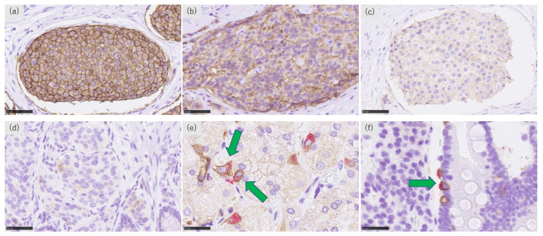
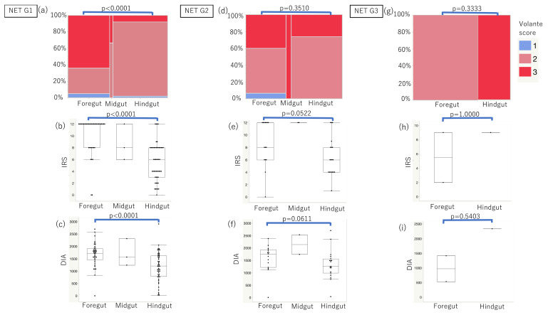
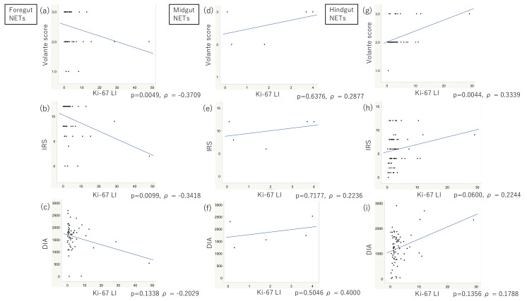
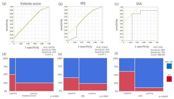
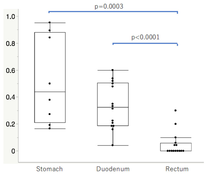
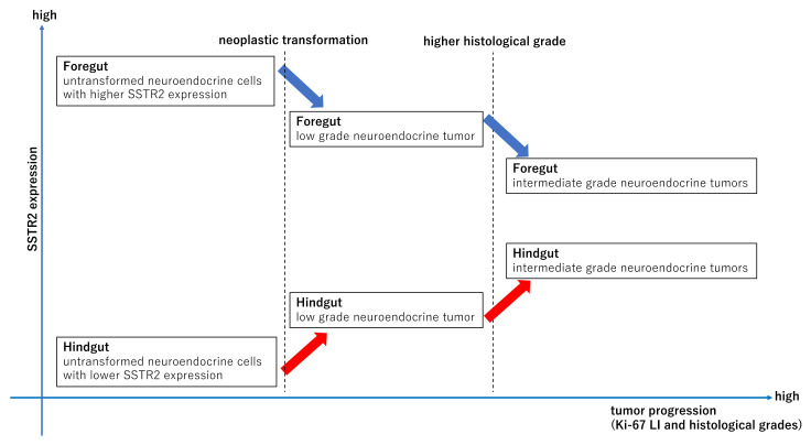
Somatostatin analogues (SSAs) are widely used to treat gastroenteropancreatic neuroendocrine tumors (GEP-NETs). Somatostatin receptor 2 (SSTR2) immunoreactivity serves as a predictive marker of the therapeutic efficacy of SSAs in pancreatic NETs. However, SSTR2 expression profiles in tumor cells and their association with the therapeutic efficacy of SSAs remains virtually unknown in gastrointestinal NETs (GI-NETs). Therefore, we evaluated the association between SSTR2 immunoreactivity and embryological origin and proliferative activity in 132 resected surgical tissues of GI-NETs. The correlation between SSAs' therapeutic efficacy and SSTR2 immunoreactivity was evaluated in 14 GI-NETs treated with SSAs. SSTR2 immunoreactivity was evaluated using Volante scores, immunoreactive scores, and digital image analysis (DIA). SSTR2 immunoreactivity was significantly negatively and positively correlated with the Ki-67 labeling index in foregut and hindgut NETs, respectively. In the normal mucosa, neuroendocrine cells in the rectum had significantly lower positive rates of SSTR2 than those in the stomach and duodenum. SSTR2 expression profiles in GI-NETs could differ by primary sites, while the difference of those between foregut and hindgut NETs might be derived from the SSTR2 status of normal neuroendocrine cell counterparts. In addition, DIA could provide a good alternative for predicting response to SSAs in evaluating SSTR2 immunoreactivity of GI-NETs.

Keywords: digital image analysis; foregut NET; hindgut NET; immunohistochemistry; neuroendocrine tumor; somatostatin receptor 2.


Conflict of interest statement

The authors declare no conflict of interest.

Figures

Figure 1
Figure 1
Representative illustrations of SSTR2 and chromogranin A immunohistochemistry. (a) Marked and circumferential, (b) moderate and incomplete, (c) weak and incomplete SSTR2 immunoreactivity in the membrane of tumor cells, (d) cytoplasmic SSTR2 immunoreactivity of tumor cells. Double immunostaining of SSTR2 and chromogranin A in the (e) stomach and (f) duodenum. Green arrows represent SSTR2 and chromogranin A double-positive neuroendocrine cells.
Figure 2
Figure 2
Correlation between SSTR2 immunoreactivity and embryonal sites of origin in NET G1, G2, and G3, respectively. (ac) In GI-NETs G1, foregut NETs demonstrated significantly higher immunoreactivity than hindgut NETs according to all scoring systems (p < 0.0001). (df) In GI-NETs G2, foregut NETs showed close to significantly higher immunoreactivity than hindgut NETs using IRSs and DIA (p = 0.0522, p = 0.0611, respectively). (gi) In GI-NETs G3, there were no significant differences of SSTR2 immunoreactivity evaluated by all scoring systems between foregut and hindgut GI-NETs.
Figure 3
Figure 3
Correlation between SSTR2 immunoreactivity and Ki-67 labeling index (LI). (ac) In the foregut NETs, SSTR2 was significantly inversely correlated with Ki-67 LI, especially when using the Volante scores and IRSs (p = 0.0049, ρ = −0.3709 and p = 0.0099, ρ = −0.3418, respectively). (df) In the midgut NETs, SSTR2 was positively correlated with Ki-67 LI, although not significantly. (gi) In the hindgut NETs, SSTR2 was positively correlated with Ki-67 LI, especially when evaluated using the Volante scores and IRSs; the former correlation was significant (p = 0.0044, ρ = 0.3339 and p = 0.0600, ρ = 0.2244, respectively).
Figure 4
Figure 4
SSTR2 immunoreactivity was evaluated by three different scoring systems and their correlation with the therapeutic efficacy of SSAs in Group 3 patients. (ac) The area under the curve (AUC) was highest when evaluated using DIA (AUC, 0.65; cut-off, 898.945; sensitivity, 80%; specificity, 75%), compared with that when using Volante scores (AUC, 0.6125; cut-off, 2; sensitivity, 90%; specificity, 25%) and IRSs (AUC, 0.5875; cutoff, 4; sensitivity, 70%; specificity, 50%). (df) Higher proportions of SD or CR were detected in positive cases than in negative cases regardless of the scoring systems used. (f) SSTR2 immunoreactivity evaluated by DIA tended to be closely associated with the therapeutic efficacy of SSAs, but not when evaluated using the Volante scores and IRSs.
Figure 5
Figure 5
SSTR2-positive rates in the normal neuroendocrine cells of gastrointestinal mucosa in Group 4 cases. SSTR2-positive rates in the stomach and duodenum were significantly higher than those in the rectum (p = 0.0003, p < 0.0001, respectively).
Figure 6
Figure 6
We hypothesized that low grade foregut NETs, derived from their normal counterparts which harbor relatively abundant SSTR2 expression, could have high SSTR2 expression, while foregut NETs with more deviation from their normal counterparts and higher histological grades could have lower SSTR2 expressions. In contrast, hindgut NETs derived from their normal counterparts which harbor intrinsically low SSTR2 expression demonstrated low SSTR2 expression, while hindgut NETs with more deviations from their normal counterparts and higher histological grades had higher SSTR2 expression.

References

    1. Taal B.G., Visser O. Epidemiology of neuroendocrine tumours. Neuroendocrinology. 2004;80:3–7. doi: 10.1159/000080731. - DOI - PubMed
    1. Ahmed M. Gastrointestinal neuroendocrine tumors in 2020. World J. Gastrointest. Oncol. 2020;12:79–807. doi: 10.4251/wjgo.v12.i8.791. - DOI - PMC - PubMed
    1. McCarthy D.M. Proton Pump Inhibitor Use, Hypergastrinemia, and Gastric Carcinoids-What Is the Relationship? Int. J. Mol. Sci. 2020;21:662. doi: 10.3390/ijms21020662. - DOI - PMC - PubMed
    1. Williams E.D., Sandler M. The classification of carcinoid tum ours. Lancet. 1963;1:238–239. doi: 10.1016/S0140-6736(63)90951-6. - DOI - PubMed
    1. Watanabe H., Yamazaki Y., Fujishima F., Izumi K., Imamura M., Hijioka S., Toriyama K., Yatabe Y., Kudo A., Motoi F., et al. O(6)-methylguanine DNA methyltransferase and glucose transporter 2 in foregut and hindgut gastrointestinal neuroendocrine neoplasms. BMC Cancer. 2020;20:1195. doi: 10.1186/s12885-020-07579-6. - DOI - PMC - PubMed

LinkOut - more resources