. 2022 Jan 25;11(3):411.
doi: 10.3390/cells11030411.

Single-Cell RNA-Seq Reveals a Crosstalk between Hyaluronan Receptor LYVE-1-Expressing Macrophages and Vascular Smooth Muscle Cells

Affiliations

Single-Cell RNA-Seq Reveals a Crosstalk between Hyaluronan Receptor LYVE-1-Expressing Macrophages and Vascular Smooth Muscle Cells

Fabienne Burger et al. Cells. .

Abstract

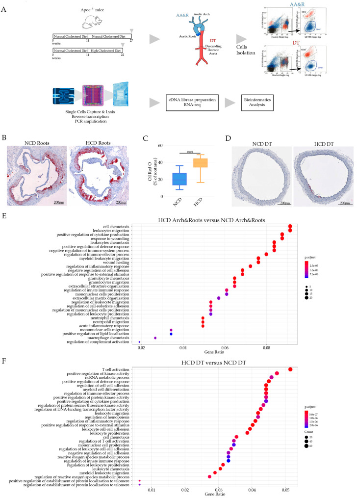
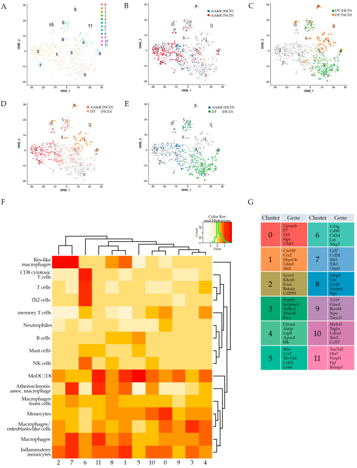
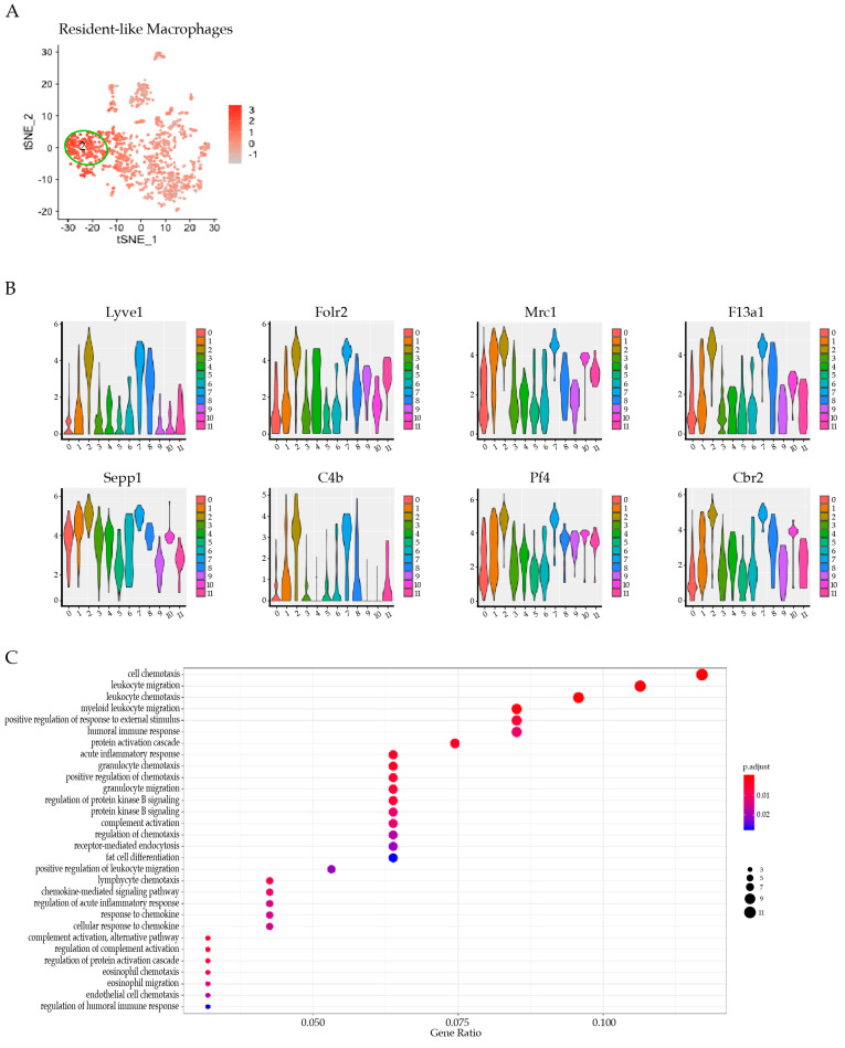
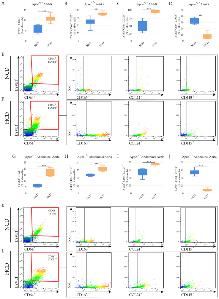
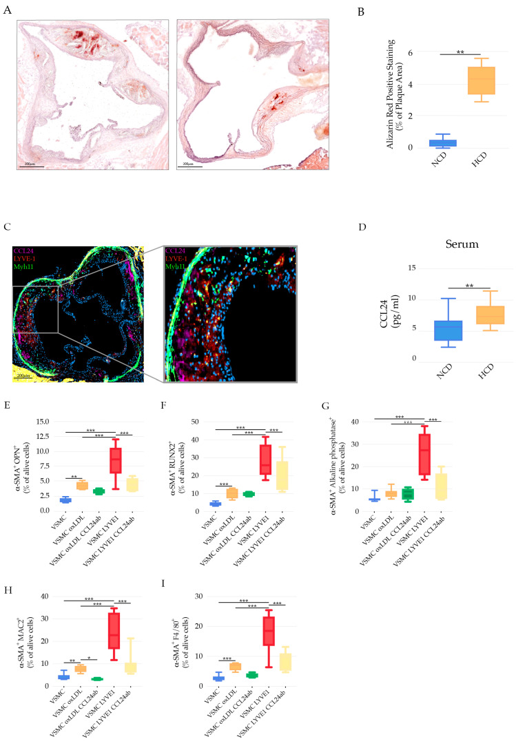
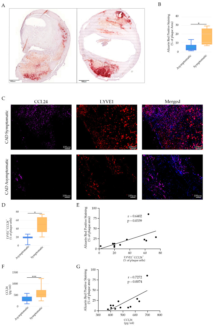
Background: Atherosclerosis is a chronic inflammatory disease where macrophages participate in the progression of the disease. However, the role of resident-like macrophages (res-like) in the atherosclerotic aorta is not completely understood. Methods: A single-cell RNA sequencing analysis of CD45+ leukocytes in the atherosclerotic aorta of apolipoprotein E-deficient (Apoe-/-) mice on a normal cholesterol diet (NCD) or a high cholesterol diet (HCD), respecting the side-to-specific predisposition to atherosclerosis, was performed. A population of res-like macrophages expressing hyaluronan receptor LYVE-1 was investigated via flow cytometry, co-culture experiments, and immunofluorescence in human atherosclerotic plaques from carotid artery disease patients (CAD). Results: We identified 12 principal leukocyte clusters with distinct atherosclerosis disease-relevant gene expression signatures. LYVE-1+ res-like macrophages, expressing a high level of CC motif chemokine ligand 24 (CCL24, eotaxin-2), expanded under hypercholesteremia in Apoe-/- mice and promoted VSMC phenotypic modulation to osteoblast/chondrocyte-like cells, ex vivo, in a CCL24-dependent manner. Moreover, the abundance of LYVE-1+CCL24+ macrophages and elevated systemic levels of CCL24 were associated with vascular calcification and CAD events. Conclusions: LYVE-1 res-like macrophages, via the secretion of CCL24, promote the transdifferentiation of VSMC to osteogenic-like cells with a possible role in vascular calcification and likely a detrimental role in atherosclerotic plaque destabilization.

Keywords: CCL24; LYVE-1; VSMC transdifferentiation; osteogenic-like cells; resident-like macrophages; vascular calcification.


Conflict of interest statement

The authors declare no conflict of interest.

Figures

Figure 1
Figure 1
(A) Experimental setting of scRNAseq of CD45+ cells of Apoe−/− on NCD versus HCD. (B) Oil Red O-stained atherosclerotic lesions of Apoe−/− mice on NCD and HCD. (C) Bar graphs represent the mean ± SEM of atherosclerotic lesion quantification in aortic roots with 6–8 mice /group and *** p < 0.001. (D) Oil Red O-stained DT aorta of Apoe−/− on NCD and HCD. (E) Bubble plot GO term enrichment analysis of cells derived from AA&R of Apoe−/− on NCD versus HCD. Dot size is proportional to the number of genes overlapping with each GO term, while the adjusted p-value is color-coded from red to blue. (F) Bubble plot GO term enrichment analysis of cells derived from DT aorta of Apoe−/− mice on NCD versus HCD. Dot size is proportional to the number of genes overlapping with each GO term, while the adjusted p-value is color-coded from red to blue.
Figure 2
Figure 2
Clusters of AA&R and DT aorta CD45+ cells of Apoe−/− on NCD and HCD; t-distributed stochastic neighbor embedding (tSNE) plot showing (A) all twelve identified clusters; (B) AA&R aorta-derived clusters of Apoe−/− mice on NCD versus HCD; (C) DT aorta-derived clusters of Apoe−/− mice on NCD versus HCD; (D) clusters of AA&R and DT aorta-derived cells of Apoe−/− mice on NCD; (E) clusters of AA&R and DT aorta-derived cells of Apoe−/− mice on HCD; (F) Heat map of the gene expression profile of 12 immune cell populations showing their immune cell population identity; (G) top 5 differentially expressed genes detected in each cluster.
Figure 3
Figure 3
Cluster 2 gene expression signature. Gene sets were overlaid on single cells on a tSNE plot to identify the cell identity of cluster 2 with an enrichment of indicated gene sets: (A) Resident-like Macrophages; and (B) top eight expressed genes of cluster 2. (C) Bubble plot GO term enrichment analysis of cells of cluster 2. Dot size is proportional to the number of genes overlapping with each GO term, while the adjusted p-value is color-coded from red to blue.
Figure 4
Figure 4
LYVE-1 res-like macrophages in mouse atherosclerotic plaques. Bar graphs represent the mean ± SEM of the percentage of LYVE1 res-like macrophages in AA&R of Apoe−/− mice on NCD versus HCD, expressed as percentage of the total live plaque cells, as defined by (A) LYVE1+CD64+; (B) LYVE1+CD64+CD163+; (C) LYVE1+CD64+CCL24+; and (D) LYVE1+CD64+CD115+, with an expression of n = 8 mice per group and *** p < 0.001. Representative dot plots show the gating strategy illustrating LYVE1+ res-like macrophage marker expression in AA&R of Apoe−/− mice on (E) NCD and (F) HCD. Bar graphs represent the mean ± SEM of the percentage of LYVE1 res-like macrophages in abdominal aortas of Apoe−/− mice on NCD versus HCD, expressed as percentage of the total live plaque cells, as defined by (G) LYVE1+CD64+; (H) LYVE1+CD64+CD163+; (I) LYVE1+CD64+CCL24+; and (J) LYVE1+CD64+CD115+, with an expression of n = 8 mice per group and *** p < 0.001. Representative dot plots illustrating the gating strategy show the LYVE1+ res-like macrophage marker expression in abdominal aortas of Apoe−/− mice on (K) NCD and (L) HCD.
Figure 5
Figure 5
The role of LYVE-1 res-like macrophages in vascular calcification. (A) Representative images of vascular calcification illustrated that Alizarin red positively stained areas in aortic root cryosections of Apoe−/− Myh11-CreERT2, ROSA26STOP-flox eYFP+/+ mice fed NCD or HCD. Scale bars: 200 μm. (B) Bar graphs represent the mean ± SEM of the percentage of Alizarin red positively stained areas in the total atherosclerotic plaque in aortic root cryosections of Apoe−/− Myh11-CreERT2, ROSA26STOP-flox eYFP+/+ mice fed NCD or HCD, n = 8 and ** p < 0.01. (C) Representative immunofluorescence staining showing LYVE1+CCL24+ cells in close proximity to VSMC (Myh11 positive cells) in atherosclerotic lesions of Apoe−/− mice on HCD; Myh11 (green), LYVE1 (red), and CCL24 (purple). (D) Bar graphs represent the mean ± SEM of systemic levels of CCL24 in Apoe−/− mice on NCD and HCD, n = 8 mice per group and ** p < 0.01. Bar graph representing the mean ± SEM of (E) α-SMA+OPN+; (F) α-SMA+RUNX2+; (G) α-SMA+ alkaline phosphatase+; (H) α-SMA+MAC2+; (I) α-SMA+F4/80+ expression in WT VSMC post-stimulation with oxLDL or co-cultured with LYVE-1 res-like macrophages in the presence or absence of CCL24 neutralizing antibodies, n = 6 and *** p < 0.001.
Figure 6
Figure 6
LYVE-1 res-like macrophages in human atherosclerotic plaques. (A) Representative images of vascular calcification illustrated by Alizarin red staining of human atherosclerotic plaques of asymptomatic and symptomatic CAD patients, respectively. Scale bars: 800 μm. (B) Bar graphs represent the mean ± SEM of the percentage of Alizarin red positively stained areas in the total atherosclerotic plaque area of asymptomatic and symptomatic CAD patients, respectively, n = 8 and * p < 0.05. (C) Representative immunofluorescence staining showing LYVE1+ cells (red) expressing CCL24+ (violet) in atherosclerotic lesions of asymptomatic and symptomatic CAD patients, respectively. (D) Bar graphs represent the mean ± SEM of LYVE1 cells expressing CCL24 in asymptomatic and symptomatic CAD patients, respectively, n = 8 and * p < 0.05. (E) Positive correlation of vascular calcification (percentage of positive Alizarin red stained areas) with percentage of LYVE1 cells expressing CCL24. (F) Bar graphs represent the mean ± SEM of systemic levels of CCL24+ in asymptomatic and symptomatic CAD patients, respectively, n = 8 and *** p < 0.001. (G) Positive correlation of vascular calcification (percentage of positive Alizarin red stained areas) with systemic levels of CCL24.

References

    1. Dickhout J.G., Basseri S., Austin R.C. Macrophage Function and Its Impact on Atherosclerotic Lesion Composition, Progression, and Stability. Arter. Thromb. Vasc. Biol. 2008;28:1413–1415. doi: 10.1161/ATVBAHA.108.169144. - DOI - PubMed
    1. Cochain C., Vafadarnejad E., Arampatzi P., Pelisek J., Winkels H., Ley K., Wolf D., Saliba A.-E., Zernecke A. Single-Cell RNA-Seq Reveals the Transcriptional Landscape and Heterogeneity of Aortic Macrophages in Murine Atherosclerosis. Circ. Res. 2018;122:1661–1674. doi: 10.1161/CIRCRESAHA.117.312509. - DOI - PubMed
    1. Zernecke A., Winkels H., Cochain C., Williams J.W., Wolf D., Soehnlein O., Robbins C.S., Monaco C., Park I., McNamara C.A., et al. Meta-Analysis of Leukocyte Diversity in Atherosclerotic Mouse Aortas. Circ. Res. 2020;127:402–426. doi: 10.1161/CIRCRESAHA.120.316903. - DOI - PMC - PubMed
    1. Tang F., Barbacioru C., Wang Y., Nordman E., Lee C., Xu N., Wang X., Bodeau J., Tuch B.B., Siddiqui A., et al. mRNA-Seq whole-transcriptome analysis of a single cell. Nat. Methods. 2009;6:377–382. doi: 10.1038/nmeth.1315. - DOI - PubMed
    1. Lim H.Y., Lim S.Y., Tan C.K., Thiam C.H., Goh C.C., Carbajo D., Chew S.H.S., See P., Chakarov S., Wang X.N., et al. Hyaluronan Receptor LYVE-1-Expressing Macrophages Maintain Arterial Tone through Hyaluronan-Mediated Regulation of Smooth Muscle Cell Collagen. Immunity. 2018;49:326–341. doi: 10.1016/j.immuni.2018.06.008. - DOI - PubMed

Publication types

MeSH terms