

Generation of an hiPSC-Derived Co-Culture System to Assess the Effects of Neuroinflammation on Blood-Brain Barrier Integrity
- PMID: 35159229
- PMCID: PMC8834542
- DOI: 10.3390/cells11030419
Generation of an hiPSC-Derived Co-Culture System to Assess the Effects of Neuroinflammation on Blood-Brain Barrier Integrity
Abstract
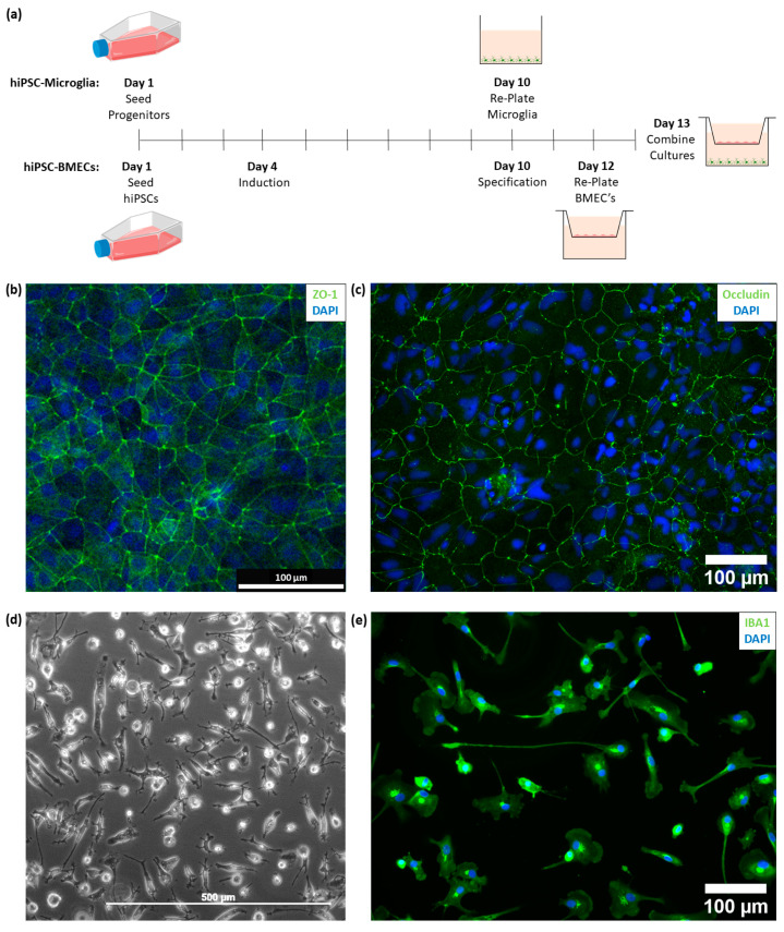
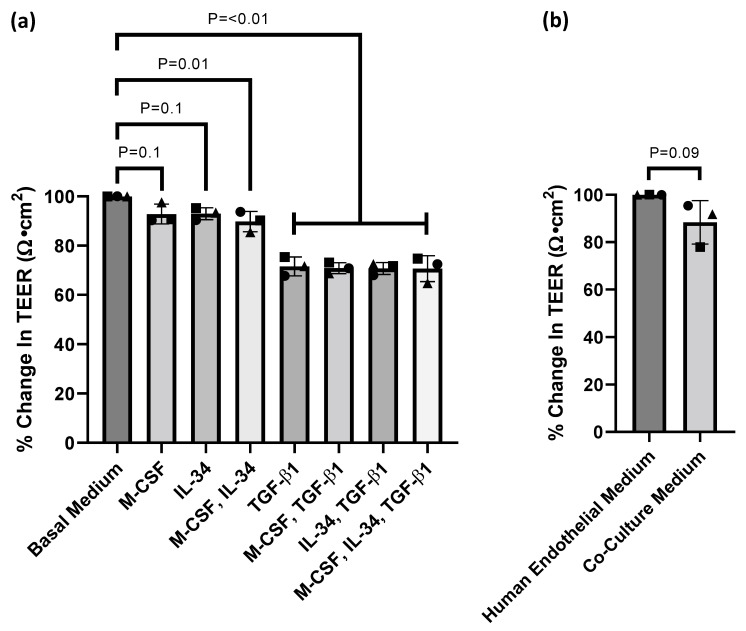
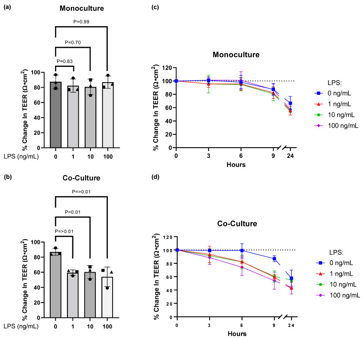
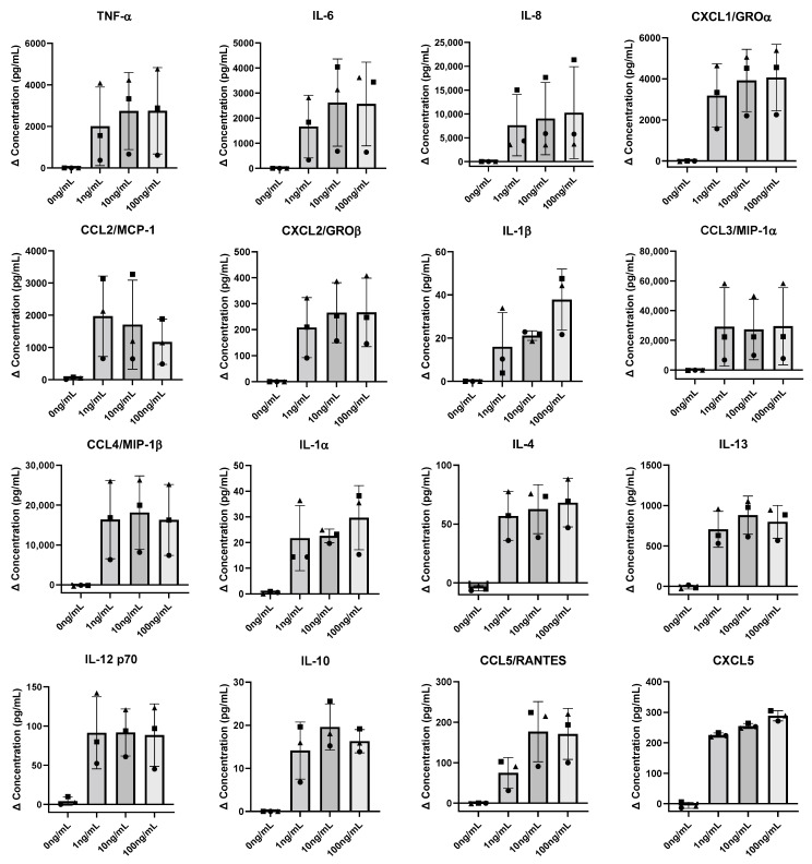
The blood-brain barrier (BBB) regulates the interaction between the highly vulnerable central nervous system (CNS) and the peripheral parts of the body. Disruption of the BBB has been associated with multiple neurological disorders, in which immune pathways in microglia are suggested to play a key role. Currently, many in vitro BBB model systems lack a physiologically relevant microglia component in order to address questions related to the mechanism of BBB integrity or the transport of molecules between the periphery and the CNS. To bridge this gap, we redefined a serum-free medium in order to allow for the successful co-culturing of human inducible pluripotent stem cell (hiPSC)-derived microglia and hiPSC-derived brain microvascular endothelial-like cells (BMECs) without influencing barrier properties as assessed by electrical resistance. We demonstrate that hiPSC-derived microglia exposed to lipopolysaccharide (LPS) weaken the barrier integrity, which is associated with the secretion of several cytokines relevant in neuroinflammation. Consequently, here we provide a simplistic humanised BBB model of neuroinflammation that can be further extended (e.g., by addition of other cell types in a more complex 3D architecture) and applied for mechanistic studies and therapeutic compound profiling.
Keywords: blood–brain barrier (BBB) integrity; brain microvascular endothelial cells (BMECs); cytokine secretion; hiPSC co-culture; microglia; neuroinflammation; neurovascular unit (NVU); transendothelial electrical resistance (TEER); transwell.
Conflict of interest statement
During the course of this study, all authors are or were full-time employees or student interns at Roche, and they may additionally hold Roche stock/stock options.
Figures
Similar articles
-
A human pluripotent stem cell-derived in vitro model of the blood-brain barrier in cerebral malaria.Fluids Barriers CNS. 2024 May 1;21(1):38. doi: 10.1186/s12987-024-00541-9. Fluids Barriers CNS. 2024. PMID: 38693577 Free PMC article.
-
Role of iPSC-derived pericytes on barrier function of iPSC-derived brain microvascular endothelial cells in 2D and 3D.Fluids Barriers CNS. 2019 Jun 6;16(1):15. doi: 10.1186/s12987-019-0136-7. Fluids Barriers CNS. 2019. PMID: 31167667 Free PMC article.
-
An hiPSC-Derived In Vitro Model of the Blood-Brain Barrier.Methods Mol Biol. 2022;2492:103-116. doi: 10.1007/978-1-0716-2289-6_5. Methods Mol Biol. 2022. PMID: 35733040
-
Neuroinflammation, Stroke, Blood-Brain Barrier Dysfunction, and Imaging Modalities.Stroke. 2022 May;53(5):1473-1486. doi: 10.1161/STROKEAHA.122.036946. Epub 2022 Apr 7. Stroke. 2022. PMID: 35387495 Free PMC article. Review.
-
Human iPSC-Derived Blood-Brain Barrier Models: Valuable Tools for Preclinical Drug Discovery and Development?Curr Protoc Stem Cell Biol. 2020 Dec;55(1):e122. doi: 10.1002/cpsc.122. Curr Protoc Stem Cell Biol. 2020. PMID: 32956578 Review.
Cited by
-
Neuroimmune Crosstalk Between the Peripheral and the Central Immune System in Amyotrophic Lateral Sclerosis.Front Aging Neurosci. 2022 May 3;14:890958. doi: 10.3389/fnagi.2022.890958. eCollection 2022. Front Aging Neurosci. 2022. PMID: 35592701 Free PMC article. Review.
-
Study of BBB Dysregulation in Neuropathogenicity Using Integrative Human Model of Blood-Brain Barrier.Front Cell Neurosci. 2022 Jun 10;16:863836. doi: 10.3389/fncel.2022.863836. eCollection 2022. Front Cell Neurosci. 2022. PMID: 35755780 Free PMC article. Review.
-
Using Human-Induced Pluripotent Stem Cell Derived Neurons on Microelectrode Arrays to Model Neurological Disease: A Review.Adv Sci (Weinh). 2023 Nov;10(33):e2301828. doi: 10.1002/advs.202301828. Epub 2023 Oct 20. Adv Sci (Weinh). 2023. PMID: 37863819 Free PMC article. Review.
References
Publication types
MeSH terms
LinkOut - more resources
Full Text Sources
Miscellaneous
