Review
. 2022 Jan 26;11(3):421.
doi: 10.3390/cells11030421.

Functional Differences between Proteasome Subtypes

Affiliations
Review

Functional Differences between Proteasome Subtypes

Joanna Abi Habib et al. Cells. .

Abstract

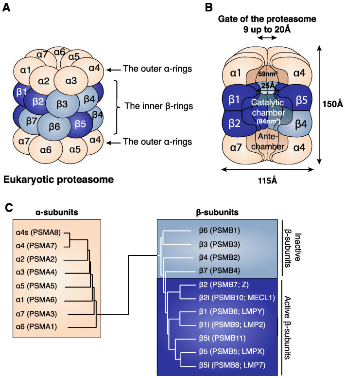
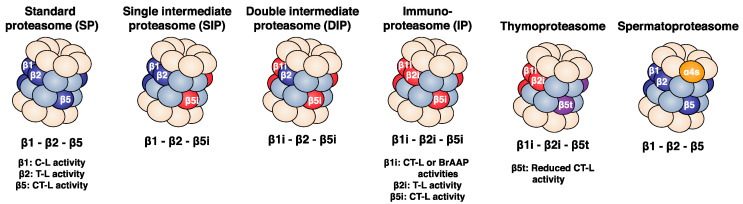
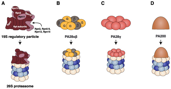
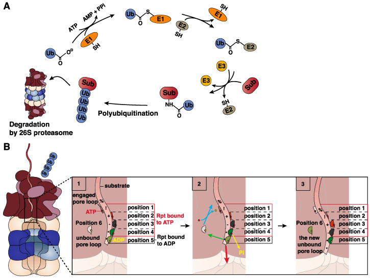
Four proteasome subtypes are commonly present in mammalian tissues: standard proteasomes, which contain the standard catalytic subunits β1, β2 and β5; immunoproteasomes containing the immuno-subunits β1i, β2i and β5i; and two intermediate proteasomes, containing a mix of standard and immuno-subunits. Recent studies revealed the expression of two tissue-specific proteasome subtypes in cortical thymic epithelial cells and in testes: thymoproteasomes and spermatoproteasomes. In this review, we describe the mechanisms that enable the ATP- and ubiquitin-dependent as well as the ATP- and ubiquitin-independent degradation of proteins by the proteasome. We focus on understanding the role of the different proteasome subtypes in maintaining protein homeostasis in normal physiological conditions through the ATP- and ubiquitin-dependent degradation of proteins. Additionally, we discuss the role of each proteasome subtype in the ATP- and ubiquitin-independent degradation of disordered proteins. We also discuss the role of the proteasome in the generation of peptides presented by MHC class I molecules and the implication of having different proteasome subtypes for the peptide repertoire presented at the cell surface. Finally, we discuss the role of the immunoproteasome in immune cells and its modulation as a potential therapy for autoimmune diseases.

Keywords: ATP- and ubiquitin-dependent degradation; ATP- and ubiquitin-independent degradation; MHC class I peptides; autoimmune diseases; proteasome subtypes; protein degradation.


Conflict of interest statement

The authors declare no conflict of interest.

Figures

Figure 1
Figure 1
(A) Structure of the eukaryotic proteasome. The identical α-rings are each composed of seven distinct α-subunits (α1–α7) (beige). The identical inner β-rings are each composed of seven distinct β-subunits (β1–β7). Only three of the seven β-subunits are catalytically active, and these are β1, β2 and β5 (dark blue). (B) Cross section of the 20S proteasome showing its internal cavities: the two antechambers of 59 nm3 and the central catalytic chamber of 84 nm3, where protein degradation takes place [9]. The length of the eukaryotic proteasome is about 150 Å and its diameter is about 115 Å [8]. The gate of the proteasome, which is delimited by the outer α-rings, has a pore size ranging from 9 to 13 Å in its closed conformation and of 20 Å in its open conformation [5,10]. (C) Relationship between the α- and β-subunits. Dendrogram showing the similarities between the eukaryotic α-subunits and between the eukaryotic β-subunits. The names of the proteasome subunits are mentioned following the common nomenclature (α and β). In parentheses we added the HUGO (human genome organization) nomenclature (PSMA and PSMB), as well as a previously common nomenclature for the catalytic subunits β1 (LMPY), β2 (Z), β5 (LMPX), β1i (LMP2), β2i (MECL1) and β5i (LMP7).
Figure 2
Figure 2
There are six proteasome subtypes that differ in their subunit composition. The standard proteasome (SP) contains the constitutive catalytic subunits β1, β2 and β5, while the immunoproteasome (IP) contains the immuno-subunits β1i, β2i and β5i. The intermediate proteasomes comprise a mixed assortment of constitutive and inducible subunits: the single intermediate proteasome (SIP) contains β1, β2 and β5i, while the double intermediate proteasome (DIP) contains β1i, β2 and β5i. The thymoproteasome contains catalytic subunit β5t, which is homologous to β5 and β5i, along with subunits β1i and β2i. Each catalytic subunit is characterized by different cleavage specificities as indicated in the lower part of the figure. Finally, the spermatoproteasome contains an α4s subunit instead of the standard α4 and expresses, in its catalytic chamber, the same assortment of catalytic subunits as the SP. Abbreviations used: caspase-like (C-L), trypsin-like (T-L), chymotrypsin-like (CT-L), and branched amino-acid preferring activity (BrAAP).
Figure 3
Figure 3
Proteasome regulators. (A) 19S regulator particle harbours two subcomplexes: the lid subcomplex contains nine subunits (Rpn3, Rpn5–9, Rpn11, Rpn12 and Rpn15; light brown) and the base subcomplex contains ten subunits (dark brown): Rpn2, Rpn1, Rpn10, Rpn13 and six distinct Rpt1-Rpt6 (regulatory particle ATPase subunits) that form the AAA+ motor of the 19S regulatory particle. The 19S regulator associates to the 20S proteasome to form the 26S proteasome. (B) The PA28αβ regulator is a ring-shaped hetero-heptameric ring composed of four PA28α subunits and three PA28β subunits. (C) The PA28γ regulator is a ring-shaped homo-heptameric ring composed of seven PA28γ subunits. (D) The PA200 regulator is a 200 kDa monomeric regulator. All four regulatory particles can bind to one or both α-rings of the 20S proteasome.
Figure 4
Figure 4
ATP- and ubiquitin-dependent degradation of proteins by the 26S proteasome. (A) Ubiquitin conjugation. Ubiquitin (Ub) conjugation requires the activity of three enzymes. E1 (a ubiquitin-activating enzyme) catalyses the ATP-dependent activation of the Ub moiety and its transfer to a conserved active cysteine on E1. Ub is then transferred to an active cysteine on E2 (a ubiquitin-conjugating enzyme). The transfer of the Ub moiety to the substrate requires the activity of an E3 enzyme (a ubiquitin ligase) that can interact with both E2 and the substrate. The resulting product is a protein–ubiquitin conjugate. Finally, the ubiquitin moiety already conjugated to the protein can undergo polymerization by the addition of ubiquitin moieties, forming a suitable tag that allows the targeting of the protein substrate for 26S proteasomal degradation. (B) Translocation of the protein substrate into the 26S proteasome. (1) Five of the six pore loops (presented here as half-circles) found in the AAA+ motor of the 19S regulatory particle align in a spiral staircase configuration entrapping the substrate. The four Rpt subunits with the pore loops at the uppermost positions are bound to an ATP molecule (red circle), and the Rpt subunit with the pore loop at the bottom position (position 5) is bound to an ADP molecule (yellow circle). The unbound pore loop occupies position 6 and is not bound to the substrate. (2) First, the Rpt subunit at position 4 hydrolyses the ATP molecule (yellow arrow), and the unbound Rpt subunit binds an ATP molecule. The newly formed Rpt-ATP subunit binds the substrate at the uppermost position as indicated by the light blue arrow, while the pore loop at position 5 disengages the substrate as indicated by the green arrow. Finally, the four Rpt subunits at positions 1–4, which are bound to the protein substrate, move downward, resulting in the translocation of the substrate (red arrow). (3) Following all these changes, the pore loop that was at position 6 occupies the uppermost position (position 1), the pore loop at position 3 occupies the penultimate position (position 4), the pore loop at position 4 occupies the bottom position (position 5), and the Rpt subunit with a pore loop at position 5 dissociates and becomes the new free pore loop at position 6 [104]. Symbols (%, !, +, -, * and °) were added on the protein substrate to aid in visualizing the translocation of the protein.
Figure 5
Figure 5
(A) Peptide bond hydrolysis. The hydroxyl group of the N-terminal threonine of the proteasome catalytic subunits attacks the carbonyl group of the peptide bond. This leads to the production of an acyl-enzyme intermediate in which the carbonyl group of the peptide fragment remains attached to the hydroxyl group of the N-terminal threonine of the proteasome by an ester link. To release the peptide fragment, a water molecule present in the catalytic chamber of the proteasome attacks the ester link between the peptide and the threonine residue, restoring the hydroxyl group of the catalytic threonine and producing the C-terminal end of the peptide. (B) Peptide splicing by the proteasome. The splicing of antigenic peptide RTK_QLYPEW derived from the gp100 is shown. Following formation of the acyl-enzyme intermediate involving the fragment RTK and the hydroxyl group of the N-terminal threonine of the proteasome, the free N-terminal amino-group of peptide QLYPEW present in the proteasome chamber attacks the acyl-enzyme intermediate, leading to the formation of the peptide RTK_QLYPEW composed of two peptide fragments originally distant in the protein.

References

    1. Zhao J., Zhai B., Gygi S.P., Goldberg A.L. mTOR inhibition activates overall protein degradation by the ubiquitin proteasome system as well as by autophagy. Proc. Natl. Acad. Sci. USA. 2015;112:15790–15797. doi: 10.1073/pnas.1521919112. - DOI - PMC - PubMed
    1. Kisselev A.F., Akopian T.N., Goldberg A.L. Range of sizes of peptide products generated during degradation of different proteins by archaeal proteasomes. J. Biol. Chem. 1998;273:1982–1989. doi: 10.1074/jbc.273.4.1982. - DOI - PubMed
    1. Vigneron N., Van den Eynde B.J. Proteasome subtypes and regulators in the processing of antigenic peptides presented by class I molecules of the major histocompatibility complex. Biomolecules. 2014;4:994–1025. doi: 10.3390/biom4040994. - DOI - PMC - PubMed
    1. Vigneron N., Abi Habib J., Van den Eynde B.J. Learning from the Proteasome How To Fine-Tune Cancer Immunotherapy. Trends Cancer. 2017;3:726–741. doi: 10.1016/j.trecan.2017.07.007. - DOI - PubMed
    1. Lowe J., Stock D., Jap B., Zwickl P., Baumeister W., Huber R. Crystal structure of the 20S proteasome from the archaeon T. acidophilum at 3.4 A resolution. Science. 1995;268:533–539. doi: 10.1126/science.7725097. - DOI - PubMed

Publication types