Review
. 2022 Jan 31;11(3):493.
doi: 10.3390/cells11030493.

Current Progress in Vascular Engineering and Its Clinical Applications

Affiliations
Review

Current Progress in Vascular Engineering and Its Clinical Applications

Hatem Jouda et al. Cells. .

Abstract

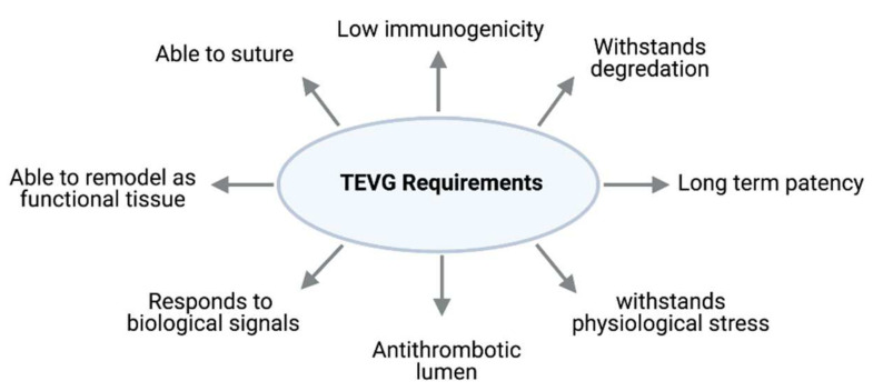
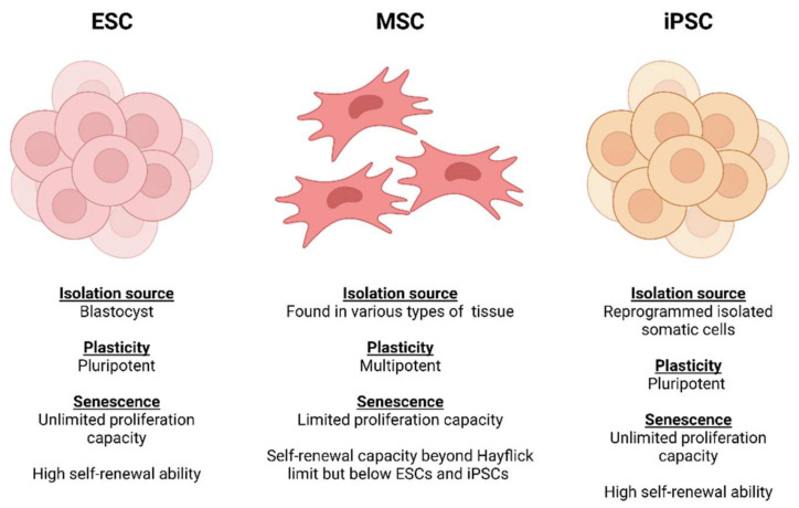
Coronary heart disease (CHD) is caused by narrowing or blockage of coronary arteries due to atherosclerosis. Coronary artery bypass grafting (CABG) is widely used for the treatment of severe CHD cases. Although autologous vessels are a preferred choice, healthy autologous vessels are not always available; hence there is a demand for tissue engineered vascular grafts (TEVGs) to be used as alternatives. However, producing clinical grade implantable TEVGs that could healthily survive in the host with long-term patency is still a great challenge. There are additional difficulties in producing small diameter (<6 mm) vascular conduits. As a result, there have not been TEVGs that are commercially available. Properties of vascular scaffolds such as tensile strength, thrombogenicity and immunogenicity are key factors that determine the biocompatibility of TEVGs. The source of vascular cells employed to produce TEVGs is a limiting factor for large-scale productions. Advanced technologies including the combined use of natural and biodegradable synthetic materials for scaffolds in conjunction with the use of mesenchyme stem cells or induced pluripotent stem cells (iPSCs) provide promising solutions for vascular tissue engineering. The aim of this review is to provide an update on various aspects in this field and the current status of TEVG clinical applications.

Keywords: induced pluripotent stem cells; ischemic heart disease; mesenchyme stem cells; tissue engineered vascular grafts; vascular tissue engineering.


Conflict of interest statement

The authors declare that they have no conflict of interest.

Figures

Figure 1
Figure 1
Structure of blood vessels. Diagram shows compositions of the three main types. From left to right: artery, vein and capillary. (Created with BioRender.com, accessed on 8 January 2022).
Figure 2
Figure 2
Vascular tissue engineering. Vascular cells can either be harvested from donors (1) or differentiated from mesenchymal stem cells (MSCs) or progenitor cells isolated from donors (2). Vascular cells can also be differentiated from pluripotent cells such as isolated embryonic stem cells (ESCs) or induced pluripotent stem cells (iPSCs) that were reprogrammed from somatic cells (e.g., dermal fibroblasts or blood monocytes) of the donor (3). A-Scaffold-based tissue-engineering: Vascular SMCs and ECs are seeded onto scaffold materials that can either be synthetic polymers or decellularised vascular scaffolds. B-Scaffold-free vascular engineering: TEVG produced via vascular cell bioprinting or rolling sheets of autologous vascular cells into a tubular structure. The constructs from A or B are then cultured ideally in a bioreactor to develop suitable properties of a TEVG for clinical implantation such as coronary artery bypass grafting. (Created with Biorender.com, accessed on 8 January 2022).
Figure 3
Figure 3
Key factors to be considered for an ideal TEVG.
Figure 4
Figure 4
Comparisons of plasticity potential, source of extraction and senescence between stem cell sources (Created with Biorender.com, accessed on 8 January 2022).
Figure 5
Figure 5
Passive seeding. Cell suspension is pipetted directly onto the lumen or exterior of the scaffold. (Created with BioRender.com, accessed on 8 January 2022).
Figure 6
Figure 6
Dynamic seeding methods. (A) Vacuum seeding: using internal or external pressure forces to drive cells into scaffolds. (B) Centrifugal/rotational seeding: using rotational force to drives cells into the scaffold. (C) Perfusion seeding: mimicking the in vivo physiological conditions and biomechanical stress of blood vessels to aid cell attachment to the scaffold. (Created with BioRender.com, accessed on 8 January 2022).
Figure 7
Figure 7
Electrostatic seeding. Scaffolds are manipulated to become positively charged substrates to attract the negatively charged regions on cell membranes for increased retention and attachment of cells. Scaffolds can be chemically modified by either cross-linking polymers in pre-polymerised solutions during the fabrication process (1), or by covering scaffold surfaces with a thin conductive layer via atomic layer deposition post-fabrication (2). The negatively charged cells can then be seeded on to the positively charged scaffold (3). Negatively charged conductors can also be employed to repel cells to increase the efficiency of cell attachment on scaffolds. (Created with BioRender.com, accessed on 8 January 2022).

References

    1. Jakobsson L., Kreuger J., Claesson-Welsh L. Building Blood Vessels—Stem Cell Models in Vascular Biology. J. Cell Biol. 2007;177:751–755. doi: 10.1083/jcb.200701146. - DOI - PMC - PubMed
    1. Ramasamy S.K. Structure and Functions of Blood Vessels and Vascular Niches in Bone. Stem Cells Int. 2017;5046953 doi: 10.1155/2017/5046953. - DOI - PMC - PubMed
    1. Hasan A., Paul A., Vrana N.E., Zhao X., Memic A., Hwang S., Dokmeci M.R., Khademhosseini A. Microfluidic Techniques for Development of 3D Vascularized Tissue. Biomaterials. 2014;35:7308–7325. doi: 10.1016/j.biomaterials.2014.04.091. - DOI - PMC - PubMed
    1. Eble J., Niland S. The Extracellular Matrix of Blood Vessels. Curr. Pharm. Des. 2009;15:1385–1400. doi: 10.2174/138161209787846757. - DOI - PubMed
    1. Fruchart J.C., Nierman M.C., Stroes E.S.G., Kastelein J.J.P., Duriez P. New Risk Factors for Atherosclerosis and Patient Risk Assessment. Circulation. 2004;109:15–19. doi: 10.1161/01.CIR.0000131513.33892.5b. - DOI - PubMed

Publication types

MeSH terms