. 2022 Jan 31;11(3):495.
doi: 10.3390/cells11030495.

Pregnant Women and Endocrine Disruptors: Role of P2X7 Receptor and Mitochondrial Alterations in Placental Cell Disorders

Affiliations

Pregnant Women and Endocrine Disruptors: Role of P2X7 Receptor and Mitochondrial Alterations in Placental Cell Disorders

Sophie Fouyet et al. Cells. .

Abstract

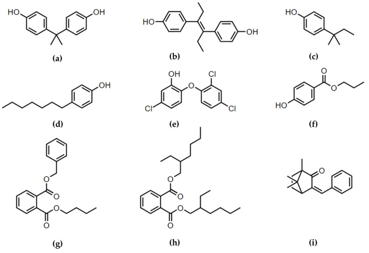
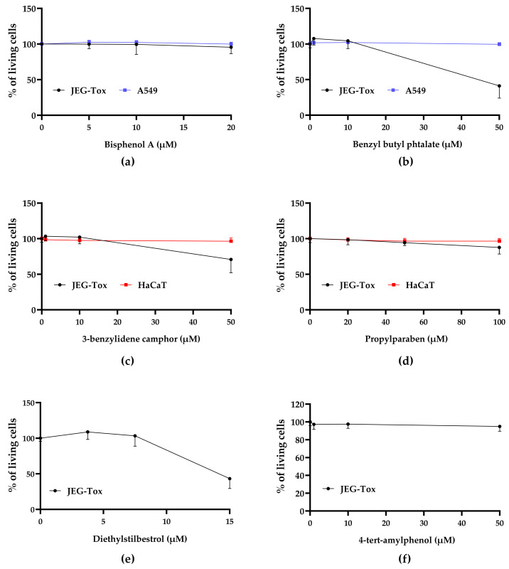
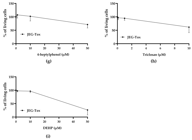
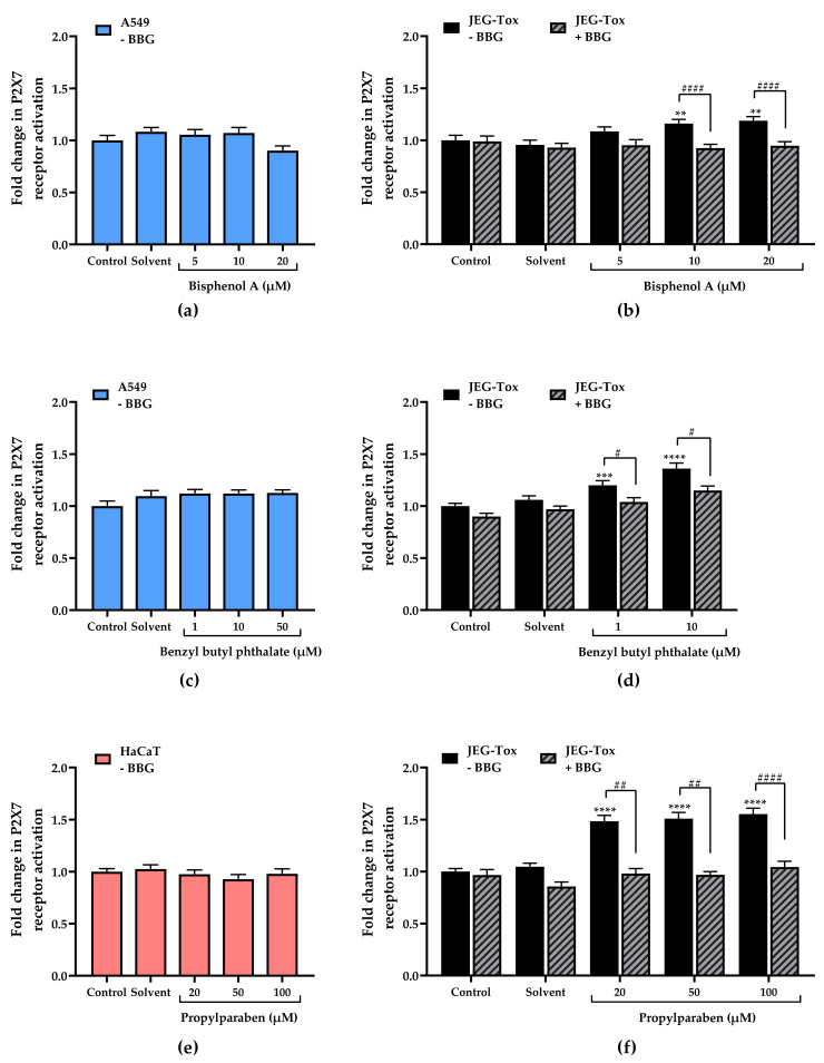
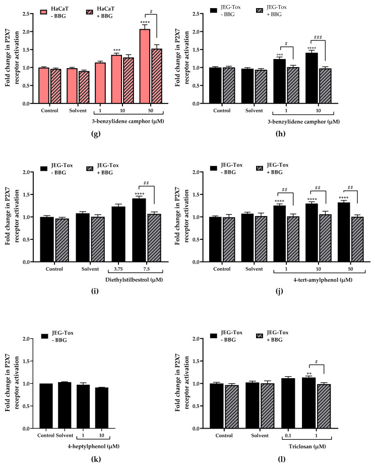
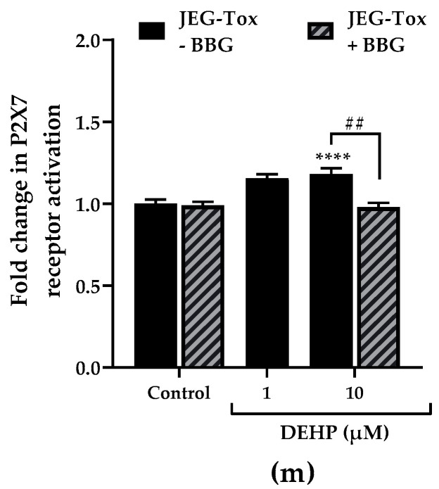
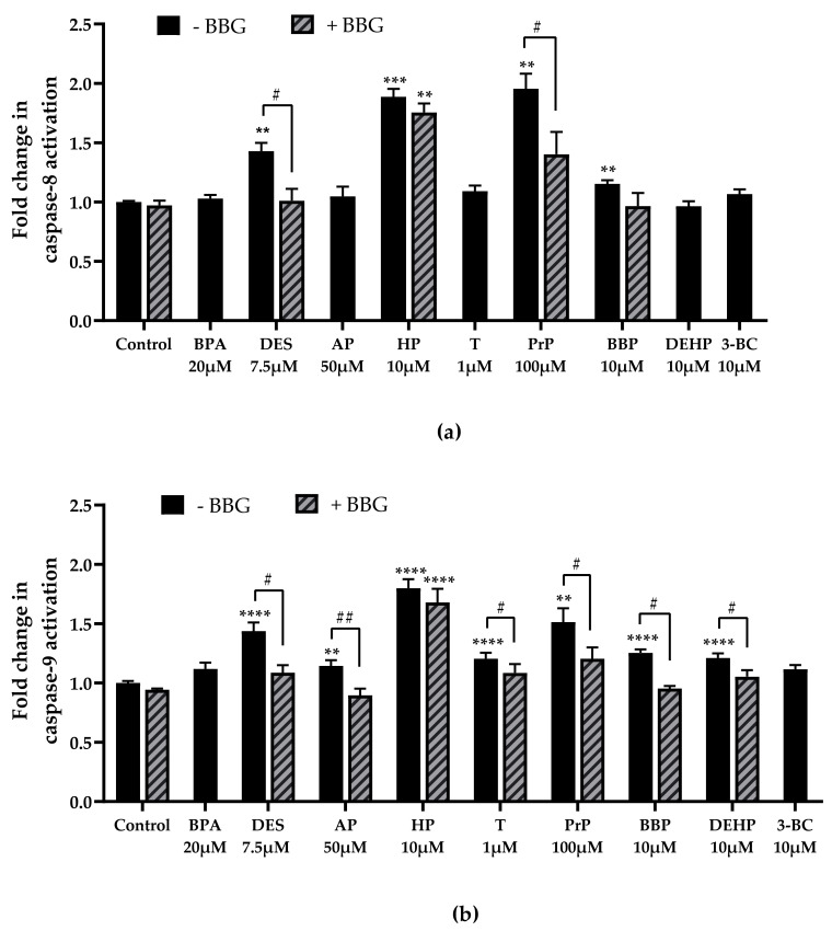
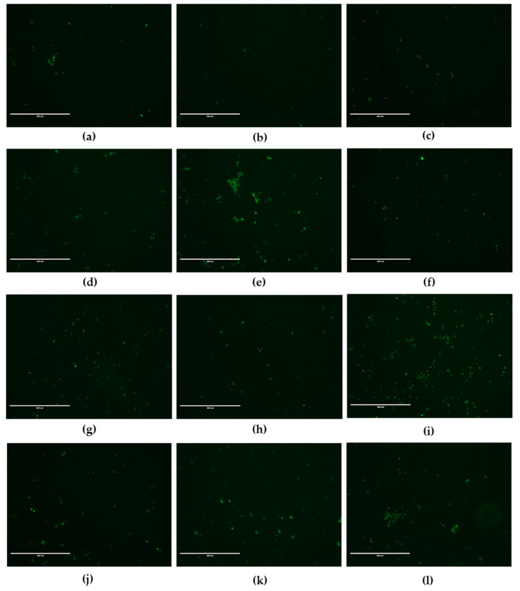
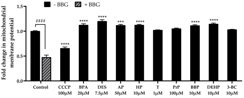
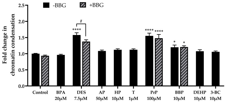
In pregnant women, the lungs, skin and placenta are exposed daily to endocrine-disrupting chemicals (EDCs). EDCs induce multiple adverse effects, not only on endocrine organs, but also on non-endocrine organs, with the P2X7 cell death receptor being potentially the common key element. Our objective was first to investigate mechanisms of EDCs toxicity in both endocrine and non-endocrine cells through P2X7 receptor activation, and second, to compare the level of activation in lung, skin and placental cells. In addition, apoptosis in placental cells was studied because the placenta is the most exposed organ to EDCs and has essential endocrine functions. A total of nine EDCs were evaluated on three human cell models. We observed that the P2X7 receptor was not activated by EDCs in lung non-endocrine cells but was activated in skin and placenta cells, with the highest activation in placenta cells. P2X7 receptor activation and apoptosis are pathways shared by all tested EDCs in endocrine placental cells. P2X7 receptor activation along with apoptosis induction could be key elements in understanding endocrine placental and skin disorders induced by EDCs.

Keywords: P2X7 receptor; apoptosis; endocrine disruptors; lung toxicity; mitochondrial alterations; placental toxicity; skin toxicity.


Conflict of interest statement

The authors declare no conflict of interest.

Figures

Figure 1
Figure 1
EDCs chemical structures: (a) bisphenol A, (b) diethylstilbestrol, (c) 4-tert-amylphenol, (d) 4-heptylphenol, (e) triclosan, (f) propylparaben, (g) benzyl butyl phthalate, (h) DEHP and (i) 3-benzylidene camphor.
Figure 2
Figure 2
Cell viability was evaluated using the neutral red assay after (a) bisphenol A and (b) benzyl butyl phthalate incubation for 72 h in A549 cells (blue bars) and JEG-Tox cells (black bars); (c) 3-benzylidene camphor and (d) propylparaben in HaCaT (red bars) and JEG-Tox cells (black bars); (e) diethylstilbestrol, (f) 4-ter-amylphenol, (g) 4-heptylphenol, (h) triclosan and (i) DEHP in JEG-Tox cells (black bars).
Figure 2
Figure 2
Cell viability was evaluated using the neutral red assay after (a) bisphenol A and (b) benzyl butyl phthalate incubation for 72 h in A549 cells (blue bars) and JEG-Tox cells (black bars); (c) 3-benzylidene camphor and (d) propylparaben in HaCaT (red bars) and JEG-Tox cells (black bars); (e) diethylstilbestrol, (f) 4-ter-amylphenol, (g) 4-heptylphenol, (h) triclosan and (i) DEHP in JEG-Tox cells (black bars).
Figure 3
Figure 3
Effects of EDCs on P2X7 receptor activation in A549 cells, HaCaT cells and JEG-Tox cells after incubation for 72 h (YO-PRO-1® assay). The cells were preincubated with either PBS (solid bars) or with Brilliant Blue G at 25 µM (hatched bars) for 15 min. The solutions were removed and A549 cells were incubated with (a) bisphenol A and (c) benzyl butyl phthalate; HaCaT cells were incubated with (e) propylparaben and (g) 3-benzylidene camphor; JEG-Tox cells were incubated with (b) bisphenol A, (d) benzyl butyl phthalate, (f) propylparaben, (h) 3-benzylidene camphor, (i) diethylstilbestrol, (j) 4-tert-amylphenol, (k) 4-heptylphenol, (l) triclosan and (m) DEHP. The significance thresholds were **** p < 0.0001, *** p < 0.001 and ** p < 0.01 compared with the control and #### p < 0.0001, ### p < 0.001, ## p < 0.01 and # p < 0.1.
Figure 3
Figure 3
Effects of EDCs on P2X7 receptor activation in A549 cells, HaCaT cells and JEG-Tox cells after incubation for 72 h (YO-PRO-1® assay). The cells were preincubated with either PBS (solid bars) or with Brilliant Blue G at 25 µM (hatched bars) for 15 min. The solutions were removed and A549 cells were incubated with (a) bisphenol A and (c) benzyl butyl phthalate; HaCaT cells were incubated with (e) propylparaben and (g) 3-benzylidene camphor; JEG-Tox cells were incubated with (b) bisphenol A, (d) benzyl butyl phthalate, (f) propylparaben, (h) 3-benzylidene camphor, (i) diethylstilbestrol, (j) 4-tert-amylphenol, (k) 4-heptylphenol, (l) triclosan and (m) DEHP. The significance thresholds were **** p < 0.0001, *** p < 0.001 and ** p < 0.01 compared with the control and #### p < 0.0001, ### p < 0.001, ## p < 0.01 and # p < 0.1.
Figure 4
Figure 4
Bisphenol A (BPA), diethylstilbestrol (DES), 4-tert-amylphenol (AP), 4-heptylphenol (HP), triclosan (T), propylparaben (PrP), benzyl butyl phthalate (BBP), DEHP and 3-benzylidene camphor (3BC) effects on caspase-8 (a) and caspase-9 (b) activity in JEG-Tox cells. The cells were preincubated with either PBS (solid bars) or BBG at 25µM (hatched bars) for 15 min. The solutions were removed and the cells were incubated with EDCs for 72 h. For clarity’s sake, EDCs abbreviations are used in Figure 4, Figure 6 and Figure 7. The significance thresholds were **** p < 0.0001, *** p < 0.001 and ** p < 0.01 compared with the control and ## p < 0.01 and # p < 0.1.
Figure 5
Figure 5
Fluorescence microscopy images of JEG-Tox cells stained for caspase-3/7 activity. After 72 h incubation with (a) control, (b) DMSO, (c) ethanol, (d) bisphenol A at 20 µM, (e) diethylstilbestrol at 7.5 µM, (f) 4-tert-amylphenol at 50 µM, (g) 4-heptylphenol at 10 µM, (h) triclosan at 1 µM, (i) propylparaben at 100 µM, (j) benzyl butyl phthalate at 10 µM, (k) DEHP at 10 µM and (l) 3-benzylidene camphor at 10 µM, the cells were stained using Caspase-3/7 Green ReadyProbes™. Representative images from three independent experiments are shown. Bar scale: 400 µm. Magnification: 10×.
Figure 6
Figure 6
Mitochondrial membrane potential was evaluated using the JC-1 assay after incubation with bisphenol A (BPA), diethylstilbestrol (DES, 4-tert-amylphenol (AP), 4-heptylphenol (HP), triclosan (T), propylparaben (PrP), benzyl butyl phthalate (BBP), DEHP and 3-benzylidene camphor (3BC) for 72 h in JEG-Tox cells. Control cells were preincubated with either PBS (solid bars) or BBG at 25µM (hatched bars) for 15 min. The solutions were removed and the cells were incubated with culture medium (control) and CCCP (positive control) for 15 min. For clarity’s sake, EDCs abbreviations are used in Figure 4, Figure 6 and Figure 7. The significance thresholds were **** p < 0.0001 and *** p < 0.001 compared with the control and #### p < 0.0001.
Figure 7
Figure 7
Chromatin condensation was assessed using the Hoechst 33342 assay after bisphenol A (BPA), diethylstilbestrol (DES, 4-tert-amylphenol (AP), 4-heptylphenol (HP), triclosan (T), propylparaben (PrP), benzyl butyl phthalate (BBP), DEHP and 3-benzylidene camphor (3BC) incubation for 72 h in JEG-Tox cells. The cells were preincubated with either PBS (solid bars) or with BBG at 25 µM (hatched bars) for 15 min. The solutions were removed and the cells were incubated with diethylstilbestrol, 4-heptylphenol, butyl benzyl phthalate (BBP) and DEHP. For clarity’s sake, EDCs abbreviations are used in Figure 4, Figure 6 and Figure 7. The significance thresholds were **** p < 0.0001 and * p < 0.1 compared with the control and # p < 0.1.

Similar articles

Cited by

References

    1. IPCS . Global Assessment of the State-of-the-Science of Endocrine Disruptor. World Health Organization; Geneva, Switzerland: 2002.
    1. Paciência I., Cavaleiro Rufo J., Silva D., Martins C., Mendes F., Farraia M., Delgado L., de Oliveira Fernandes E., Padrão P., Moreira P., et al. Exposure to indoor endocrine-disrupting chemicals and childhood asthma and obesity. Allergy. 2019;74:1277–1291. doi: 10.1111/all.13740. - DOI - PubMed
    1. Ju Q., Zouboulis C.C. Endocrine-disrupting chemicals and skin manifestations. Rev. Endocr. Metab. Disord. 2016;17:449–457. doi: 10.1007/s11154-016-9371-2. - DOI - PubMed
    1. Bamigboye A.A., Morris J. Oestrogen supplementation, mainly diethylstilbestrol, for preventing miscarriages and other adverse pregnancy outcomes. Cochrane Database Syst. Rev. 2003;3:CD004353. doi: 10.1002/14651858.CD004353. - DOI - PMC - PubMed
    1. Chen X., Chen M., Xu B., Tang R., Han X., Qin Y., Xu B., Hang B., Mao Z., Huo W., et al. Parental phenols exposure and spontaneous abortion in Chinese population residing in the middle and lower reaches of the Yangtze River. Chemosphere. 2013;93:217–222. doi: 10.1016/j.chemosphere.2013.04.067. - DOI - PubMed

Substances

LinkOut - more resources