. 2022 Feb 1;11(3):504.
doi: 10.3390/cells11030504.

Intestinal Microbiota Remodeling Protects Mice from Western Diet-Induced Brain Inflammation and Cognitive Decline

Affiliations

Intestinal Microbiota Remodeling Protects Mice from Western Diet-Induced Brain Inflammation and Cognitive Decline

Prasant Kumar Jena et al. Cells. .

Abstract

It has been shown that the Western diet (WD) induces systemic inflammation and cognitive decline. Moreover, probiotic supplementation and antibiotic treatment reduce diet-induced hepatic inflammation. The current study examines whether shaping the gut microbes by Bifidobacterium infantis (B. infantis) supplementation and antibiotic treatment reduce diet-induced brain inflammation and improve neuroplasticity. Furthermore, the significance of bile acid (BA) signaling in regulating brain inflammation was studied. Mice were fed a control diet (CD) or WD for seven months. B. infantis was supplemented to WD-fed mice to study brain inflammation, lipid, metabolomes, and neuroplasticity measured by long-term potentiation (LTP). Broad-spectrum coverage antibiotics and cholestyramine treatments were performed to study the impact of WD-associated gut microbes and BA in brain inflammation. Probiotic B. infantis supplementation inhibited diet-induced brain inflammation by reducing IL6, TNFα, and CD11b levels. B. infantis improved LTP and increased brain PSD95 and BDNF levels, which were reduced due to WD intake. Additionally, B. infantis reduced cecal cholesterol, brain ceramide and enhanced saturated fatty acids. Moreover, antibiotic treatment, as well as cholestyramine, diminished WD-induced brain inflammatory signaling. Our findings support the theory that intestinal microbiota remodeling by B. infantis reduces brain inflammation, activates BA receptor signaling, and improves neuroplasticity.

Keywords: Bifidobacterium infantis; bile acid receptor; brain inflammation; gut microbiota; metabolomics; neuroplasticity.


Conflict of interest statement

The authors declare no conflict of interest.

Figures
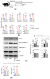
Figure 1
Figure 1
Probiotic B. infantis reduced brain inflammation signaling in WD-fed mice. (a) Experimental schema. (b) The hippocampal mRNA levels of inflammatory signaling genes of CD-fed (n = 6), WD-fed (n = 6), and WD-fed mice supplemented with B. infantis (n = 4). (c) Western blot of proteins related to inflammatory signaling. (d) Brain homogenate concentration of IL1β and TNFα by ELISA (n = 3). Data are expressed as means ± SD. One-Way ANOVA multiple comparisons Tukey t-test, * p < 0.05, ** p < 0.01, *** p < 0.001. CD-fed mice vs. WD-fed mice; WD-fed mice vs. B. infantis supplemented mice.
Figure 2
Figure 2
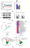
B. infantis treatment improved long-term potentiation (LTP) and synaptic deficits as well as an altered brain lipid profile: (a) Scatter plot indicates high-frequency stimulation-induced LTP, and the bar graph shows LTP calculated by averaging the change in fEPSP slope apparent between 50 and 60 min after high-frequency stimulation (n = 3, 9 slices per brain); all data is presented as the percent change in fEPSP slope means ± SEM from baseline; (b) Western blot data shows protein levels in the brains (CD, n = 6; WD, n = 6; WD + B. infantis, n = 4); (c) sPLS-DA based analysis of brain lipidomics; (d) Heatmap analysis shows a mean value of pick intensity of the top 23 lipids changed in each experimental group; (e) Volcano plots represent the brain lipidomics profile between WD vs. CD and WD + B. infantis vs. WD. The red color represents the fold changes of >2 with a p-value < 0.05, (n = 4). Data is mean ± SD. * p < 0.05, ** p < 0.01, *** p < 0.001.
Figure 3
Figure 3
Untargeted metabolomics study of cecal content. Mice were fed a WD and treated with or without B. infantis: (a) sPLS-DA analysis of cecal metabolites clustered differently; (b) Volcano plots represent the cecal metabolomics profile between WD vs. CD (n = 4) and WD + B. infantis vs. WD (n = 4). The red color represents the fold changes of > 2 with a p-value < 0.05; (c) ChemRICH metabolite set enrichment statistics plot. The node color shows increased (red) or decreased (blue) metabolite sets. Only enrichment clusters are shown significantly different at p < 0.05 (The node sizes represent the total number of metabolites in each cluster set); (d) The pathway impact and (e) pathway analysis impacts were shown between WD vs. CD (n = 4) and WD + B. infantis vs. WD (n = 4).
Figure 4
Figure 4
Probiotic B. infantis prevented gut dysbiosis induced by WD: (a) Relative abundance of cecal microbiota at the phylum level; (b) The ratio between phylum Firmicutes and Bacteroidetes; (c) Relative abundance of cecal microbiota at the family level; (d) Relative abundance of cecal microbiota at genus level in control diet (CD) and WD fed mice treated with and without B. infantis for 2 months (CD, n = 6; WD, n = 6; WD + B. infantis, n = 4). Data are expressed as mean ± SD. * p < 0.05, ** p < 0.01, *** p < 0.001, CD-fed mice compared with WD-fed mice and WD-fed mice compared with B. infantis treated mice.
Figure 5
Figure 5
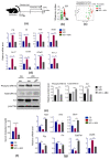
Microbiota depletion by antibiotics abrogated the inflammation induced by WD: (a) Experimental schema; (b) Shannon diversity index; (c) PCoA plot shows unweighted unifrac distance of fecal microbiota after 16S sequencing; (d) Relative mRNA level of inflammatory signaling genes in brain (CD, n = 6; WD, n = 6; WD with ABX, n = 4); (e) Western blot of inflammation signaling-related proteins in the brain of all experimental groups; (f) Serum cholesterol level and relative mRNA level of bile acid signaling genes in the brain of all experimental groups; (g) Relative mRNA level of bile acid receptor signaling genes in the brain. Data are expressed as mean ± SD. * p < 0.05, ** p < 0.01, *** p < 0.001, CD-fed compared with WD-fed mice, WD-fed mice compared with ABX (cocktail of ampicillin, neomycin, vancomycin, and metronidazole) treated mice.
Figure 6
Figure 6
Probiotic B. infantis improved bile acid receptor signaling in the brain reduced by WD: (a) Bile acid signaling genes of CD (n = 6), WD (n = 6), and WD fed with B. infantis (n = 4) in mouse brains at the mRNA level; (b) The protein level of G-protein coupled bile acid receptor 1 (Gpbar1) in the brain; (c) Secondary to primary BA ratio, conjugated to unconjugated BA ratio, and serum bile acid level of CD, WD, and WD fed with B. infantis mice. Data are expressed as means ± SD. One-Way ANOVA multiple comparisons Tukey t-test, * p < 0.05, ** p < 0.01, *** p < 0.001. CD-fed mice compared with WD-fed mice and WD-fed mice compared with B. infantis treated mice.
Figure 7
Figure 7
Effect of bile acid depletion by cholestyramine on brain inflammation and bile acid receptor signaling: (a) Experimental schema; (b) Relative mRNA level of inflammatory signaling genes in the brain (CD, n = 6; WD, n = 6; WD + cholestyramine n = 4); (c) Relative mRNA level of bile acid receptor signaling genes in the brain of all experimental groups. Data are expressed as mean ± SD. * p < 0.05, ** p < 0.01, *** p < 0.001, CD-fed compared with WD-fed mice, WD-fed mice compared with cholestyramine treated mice.

References

    1. Noble E.E., Hsu T.M., Kanoski S.E. Gut to Brain Dysbiosis: Mechanisms Linking Western Diet Consumption, the Microbiome, and Cognitive Impairment. Front. Behav. Neurosci. 2017;11:9. doi: 10.3389/fnbeh.2017.00009. - DOI - PMC - PubMed
    1. Jena P.K., Sheng L., Di Lucente J., Jin L.W., Maezawa I., Wan Y.Y. Dysregulated bile acid synthesis and dysbiosis are implicated in Western diet-induced systemic inflammation, microglial activation, and reduced neuroplasticity. FASEB J. 2018;32:2866–2877. doi: 10.1096/fj.201700984RR. - DOI - PMC - PubMed
    1. Kanoski S.E., Davidson T.L. Different patterns of memory impairments accompany short- and longer-term maintenance on a high-energy diet. J. Exp. Psychol. Anim. Behav. Processes. 2010;36:313–319. doi: 10.1037/a0017228. - DOI - PubMed
    1. Murray A.J., Knight N.S., Cochlin L.E., McAleese S., Deacon R.M., Rawlins J.N., Clarke K. Deterioration of physical performance and cognitive function in rats with short-term high-fat feeding. FASEB J. 2009;23:4353–4360. doi: 10.1096/fj.09-139691. - DOI - PubMed
    1. Khazen T., Hatoum O.A., Ferreira G., Maroun M. Acute exposure to a high-fat diet in juvenile male rats disrupts hippocampal-dependent memory and plasticity through glucocorticoids. Sci. Rep. 2019;9:12270. doi: 10.1038/s41598-019-48800-2. - DOI - PMC - PubMed

Publication types