. 2022 Feb 5;11(3):558.
doi: 10.3390/cells11030558.

Induction of Survival of Motor Neuron (SMN) Protein Deficiency in Spinal Astrocytes by Small Interfering RNA as an In Vitro Model of Spinal Muscular Atrophy

Affiliations

Induction of Survival of Motor Neuron (SMN) Protein Deficiency in Spinal Astrocytes by Small Interfering RNA as an In Vitro Model of Spinal Muscular Atrophy

Markus Leo et al. Cells. .

Abstract

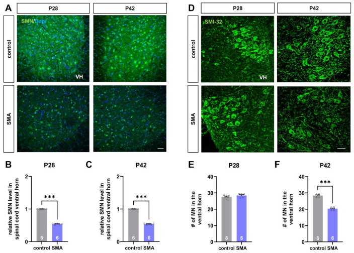
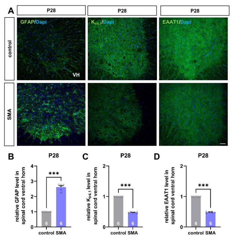
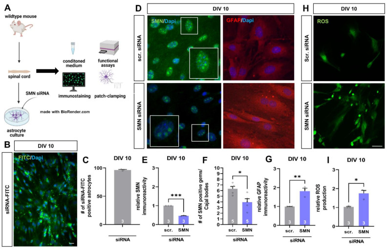
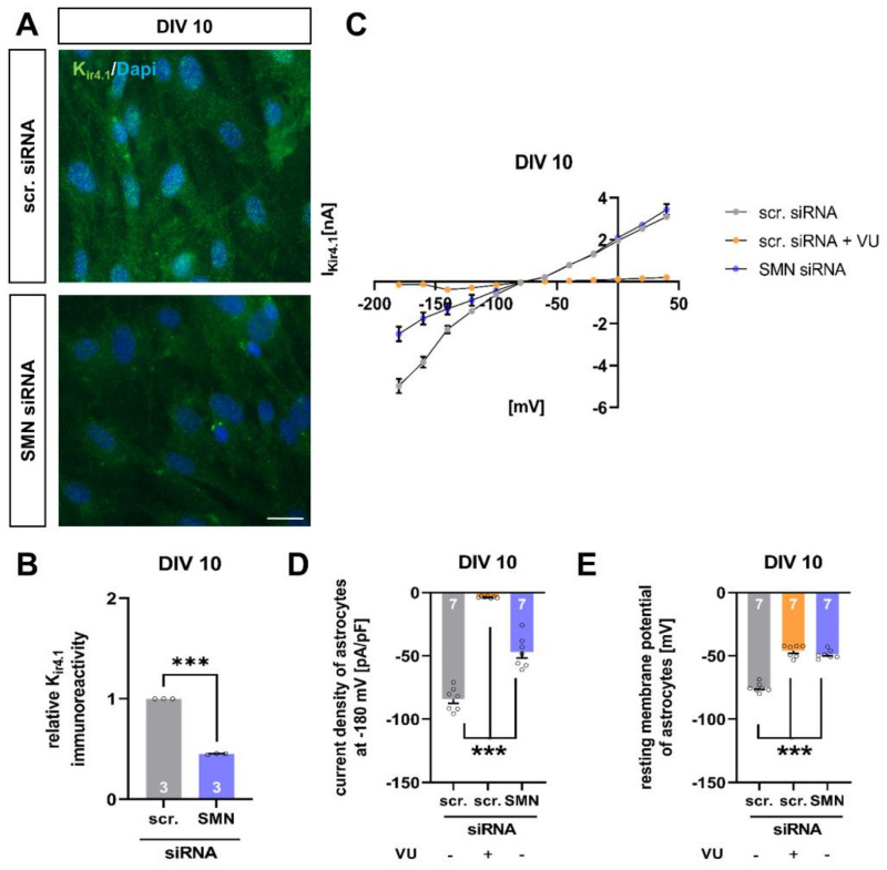
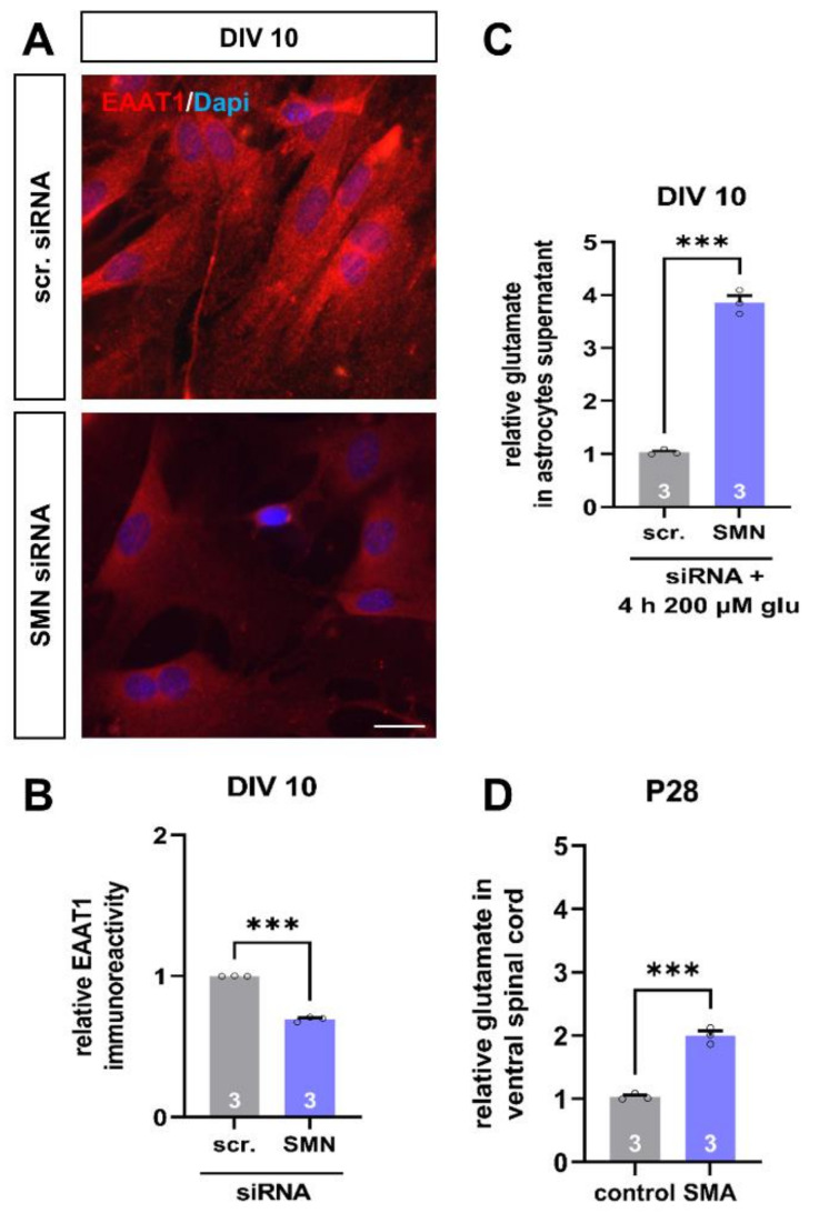
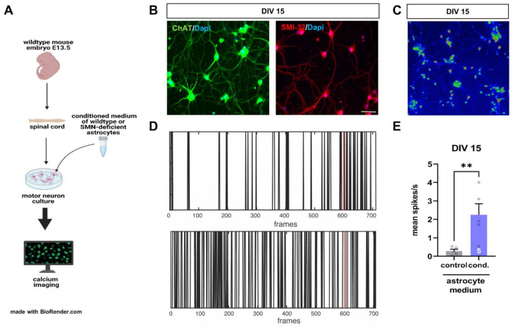
Spinal muscular atrophy (SMA) is a motor neuron disorder leading to progressive loss of ventral horn neurons resulting in muscle wasting. Here we investigate the contribution of spinal astrocytes to the pathogenesis of late-onset SMA forms using a mouse model. Furthermore, we generated SMA-like astrocytes using survival of motor neuron (SMN) siRNA transfection techniques. In the SMA mouse model, the activation of spinal astrocytes and the reduction of the inward rectifier potassium channel Kir4.1 and excitatory amino acid transporter 1 (EAAT1) were observed at postnatal day (P) 28, preceding the loss of spinal motor neurons appearing earliest at P42. Using SMA-like astrocytes, we could mimic the modulation of spinal astrocytes of the mouse model in a dish and perform electrophysiological assessments and functional assays. In SMA-like astrocytes, glutamate uptake was diminished due to a reduction in EAAT1. Furthermore, patch-clamp measurements revealed reduced potassium uptake into astrocytes with membrane depolarization. Additionally, exposure of healthy spinal motor neurons to a conditioned medium of SMA-like astrocytes resulted in increased firing frequency. These data demonstrate spinal astrocytes' crucial role in the late-onset SMA forms' pathogenesis.

Keywords: SMA; SMN; astrocytes; glia–neuron interaction; neuromuscular disorders; siRNA; spinal cord; spinal muscular atrophy; survival of motor neuron.


Conflict of interest statement

The authors declare no conflict of interest.

Figures

Figure 1
Figure 1
Deficiency of SMN protein and loss of spinal motor neurons in a mouse model of late-onset SMA. (A) Immunostaining of SMN protein (green) in spinal cord slices of control and SMA mice at P28 and P42. Nuclear DNA was stained with Dapi (blue). (B,C) SMN protein level was reduced in the spinal cord ventral horn of SMA mice at P28 and P42, compared to control mice at the same age (***, p < 0.001). (D) Immunostaining of spinal motor neurons (SMI-32, green) in spinal cord slices of control and SMA mice at P28 and P42. (E,F) The number of spinal motor neurons was reduced in SMA mice at P42, compared to control animals (***, p < 0.001). No loss of spinal motor neurons was observed at P28 (p > 0.05). VH = ventral horn. n = 6 animals (see bars). Three slices per spinal cord were investigated. Each data point reflects the mean of those three spinal cord slices. Scale bar: A = 20 µm; B = 50 µm.
Figure 2
Figure 2
Spinal astrocytes proteins are altered in a mouse model of late-onset SMA before the first loss of motor neurons. (A) Immunostaining of astrocyte-specific proteins such as GFAP (green), Kir4.1 (green), and EAAT1 (green) in spinal cord slices of SMA and control mice at P28. Nucleic DNA was stained with Dapi (blue). (B) The relative GFAP level in SMA mice spinal cord ventral horns was increased compared to control mice (***, p < 0.001). (C,D) The relative level of Kir4.1 and EAAT1 was reduced in SMA mice, compared to control animals (***, p < 0.001). VH = ventral horn. n = 6 animals (see bars). Three slices per spinal cord were investigated. Each data point reflects the mean of those three spinal cord slices. Scale bar: 20 µm.
Figure 3
Figure 3
SMN-deficient spinal astrocytes can be generated by SMN siRNA in vitro, showing similar protein alteration as spinal astrocytes in the late-onset SMA mouse model. (A) Schematic drawing of the generation of SMA-deficient spinal astrocyte cultures by SMN siRNA and performed experiments. (B) Transfection of wild-type astrocyte cultures with scrambled siRNA-FITC (green) by magnetic transfection method. Nuclear DNA was stained with Dapi (blue). (C) Cultured wild-type astrocytes were successfully transfected with siRNA-FITC using the magnetic transfection method. (D) Immunostaining of wild-type astrocyte cultures transfected with scrambled (scr.) or SMN siRNA against SMN protein (green) and GFAP (red) at DIV 10. In addition, nuclear DNA was stained with Dapi (blue). (E) Transfection of wild-type spinal astrocytes with SMN siRNA resulted in a reduction of SMN protein similar to the level observed in the juvenile SMA type III mouse model (***, p < 0.001). (F) SMN positive gems/Cajal bodies in the nucleus of astrocytes were reduced by transfection with SMN siRNA (*, p < 0.05). Furthermore, an increased number of GFAP-positive aggregate-like structures were observed in SMN-deficient astrocytes (white arrows). (G) SMN-deficiency in spinal astrocytes induced by SMN siRNA resulted in increased GFAP level (**, p < 0.01). (H) Staining of ROS production (green) in wild-type astrocytes transfected with scrambled or SMN siRNA at DIV 10. (I) SMN-deficient astrocytes showed an increased production of ROS compared to astrocytes transfected with scrambled siRNA (*, p < 0.05). n = 3–5 independent experiments (see bars). For each experiment > 50 cells were analyzed. Scale bar: B = 50 µm; F = 20 µm.
Figure 4
Figure 4
SMN-deficient spinal astrocytes show a reduction in Kir4.1 protein level and modulation in electrophysiological properties. (A) Immunostaining of wild-type astrocyte cultures transfected with scrambled (scr.) or SMN siRNA against Kir4.1 protein (green) at DIV 10. Nuclear DNA was stained with Dapi (blue). (B) SMN-deficient spinal astrocytes showed reduced protein levels of Kir4.1, compared to astrocytes transfected with scrambled siRNA (***, p < 0.001). (C) Raw trace of current–voltage (IV) curve of Kir4.1 channel protein in cultured spinal astrocytes transfected with scrambled or SMN siRNA or scrambled siRNA + Kir4.1 inhibitor VU. SMN-deficient astrocytes showed reduced current as a sign of reduced potassium uptake into spinal astrocytes. A similar effect was observed when VU inhibited the Kir4.1 function. (D) Current density of Kir4.1 was reduced in SMN-deficient spinal astrocytes, compared to astrocytes transfected with scrambled siRNA (***, p < 0.001). When Kir4.1 function was inhibited in wild-type astrocytes exposed to scrambled siRNA, the current density was reduced (***, p < 0.001). (E) Astrocytes transfected with SMN siRNA showed depolarized resting membrane potential (***, p < 0.001). A similar result was observed when Kir4.1 function was inhibited by VU (***, p < 0.001). n = 3 independent experiments for immunostaining (see bars). For each experiment > 50 cells were analyzed. n = 7 cells per electrophysiological measurement (see bars). Scale bar: 50 µm.
Figure 5
Figure 5
SMN-deficient spinal astrocytes show a reduction in EAAT1 protein level and reduced uptake of glutamate. (A) Immunostaining of wild-type astrocyte cultures transfected with scrambled (scr.) or SMN siRNA against EAAT1 protein (green) at DIV 10. In addition, nuclear DNA was stained with Dapi (blue). (B) SMN-deficient spinal astrocytes showed reduced protein levels of EAAT1, compared to astrocytes transfected with scrambled siRNA (***, p < 0.001). (C) When astrocytes were exposed to 200 µM of glutamate (glu) for 4 h, SMN-deficient astrocytes showed reduced uptake of the transmitter, compared to astrocytes transfected with scrambled siRNA (***, p < 0.001). (D) Ventral horn spinal cord tissue of juvenile SMA type III mouse model showed an increased glutamate level at P28, compared to control animals (***, p < 0.001). n = 3 independent experiments for immunostaining (see bars). For each experiment > 50 cells were analyzed. n = 3 independent experiments per functional measurement (see bars). Glutamate measurements of spinal cord tissue were from three individual animals. Scale bar: 50 µm.
Figure 6
Figure 6
Conditioned medium of SMN-deficient astrocytes leads to hyperactivity of cultured spinal motor neurons of wild-type mice. (A) Schematic drawing of experimental design. Motor neurons were isolated from E13.5 embryos of wild-type mice. Cultured motor neurons were exposed to a conditioned medium of astrocytes transfected with scrambled or SMN siRNA for 24 h. Afterward, calcium imaging measurements were performed. (B) Immunostaining of cultured motor neurons at DIV 15. Neurons expressed typical markers of motor neurons such as ChAT (green) or SMN-32 (red). In addition, nuclear DNA was stained with Dapi (blue). (C) Image with Fluo-4 AM stained motoneurons at DIV 15 in LUT (16 colors). (D) Image of spiking events over the recording period. The red line indicates the application of potassium pulse (60 mM KCl). (E) Motor neurons exposed to a conditioned medium of SMN-deficient astrocytes showed an increased number in their spiking frequency (**, p < 0.01). n = 5 motor neurons measured per experiment (see bars). Scale bar: 20 µm.

Similar articles

Cited by

References

    1. Eroglu C., Barres B.A. Regulation of synaptic connectivity by glia. Nature. 2010;468:223–231. doi: 10.1038/nature09612. - DOI - PMC - PubMed
    1. Liddelow S.A., Guttenplan K.A., Clarke L.E., Bennett F.C., Bohlen C.J., Schirmer L., Bennett M.L., Munch A.E., Chung W.S., Peterson T.C., et al. Neurotoxic reactive astrocytes are induced by activated microglia. Nature. 2017;541:481–487. doi: 10.1038/nature21029. - DOI - PMC - PubMed
    1. Barbour B., Brew H., Attwell D. Electrogenic glutamate uptake in glial cells is activated by intracellular potassium. Nature. 1988;335:433–435. doi: 10.1038/335433a0. - DOI - PubMed
    1. Gee J.R., Keller J.N. Astrocytes: Regulation of brain homeostasis via apolipoprotein E. Int. J. Biochem. Cell Biol. 2005;37:1145–1150. doi: 10.1016/j.biocel.2004.10.004. - DOI - PubMed
    1. Sasaki S., Komori T., Iwata M. Excitatory amino acid transporter 1 and 2 immunoreactivity in the spinal cord in amyotrophic lateral sclerosis. Acta Neuropathol. 2000;100:138–144. doi: 10.1007/s004019900159. - DOI - PubMed

Publication types

Substances

LinkOut - more resources